카테고리 보관물: JNOPTIC

JNO-R1(B)

JNOPTIC Filter JNO-R1(B)

Download Catalog_JNO-R1(B)

– Filter set for Texas Red / mCherry / AlexaFluor 594
– Chroma Filter Model No. : 39010

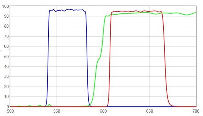
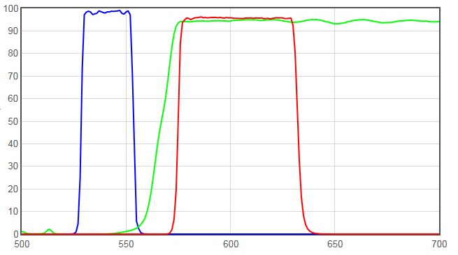
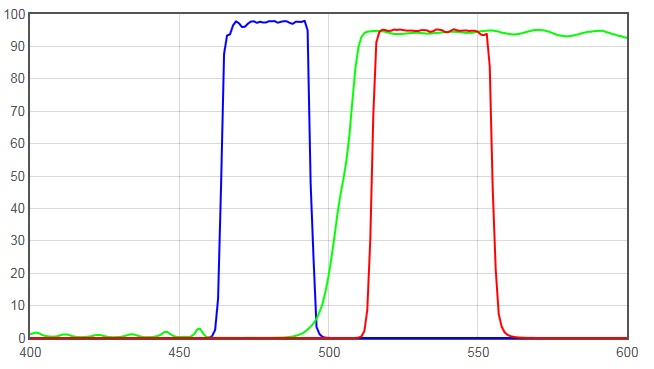
JNO-R1(B) Spectrum

JNO-R1(B)
JNO-R1(B)
FiltersType
AT560/40xEX
AT600DCBS
AT635/60mEM

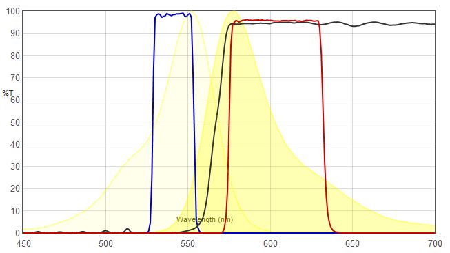
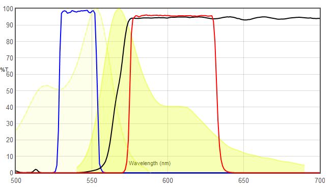
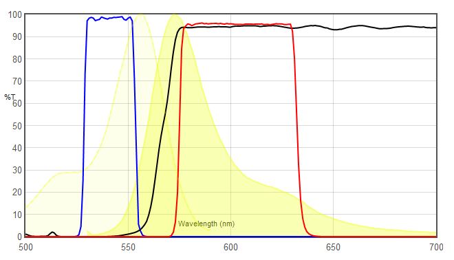
주요 형광 색소(Fluorochromes)와 스펙트럼 비교

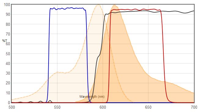
Texas Red VS JNO-R1(B)
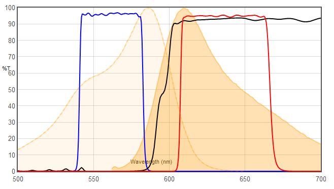
mCherry VS JNO-R1(B)
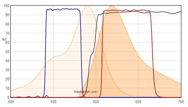
Alexa Fluor 594 VS JNO-R1(B)

JNO-R1(B)필터에 적합한 형광색소


JNO-R1(B) Spec

  • Typical Application(s) : Widefield Microscopy
  • Coating : Sputter/Hard Coated
  • Round excitation and emission filters mounted in anodized aluminum rings 2.3mm thick, up to 25mm in diameter (for standard sizes)
  • Rectangular dichroics up to 26x38mm, 1mm thick, to fit standard microscope manufacturer filter cubes (for standard sizes)

*** For filters larger than 25mm and dichroics larger than 26x38mm, please contact us for pricing.

JNO-G(B)

JNOPTIC Filter JNO-G(B)

Download Catalog _ JNO-G(B)

– Filter set for TRITC / Cy3 / TagRFP / AlexaFluor546
– Chroma Filter Model No. : 39004

JNO-G(B) Spectrum

JNO-G(B)
Standard Green Excitaion filter (Bans Pass type)
FiltersType
AT540/25xEX
AT565DCBS
AT605/55mEM

주요 형광 색소(Fluorochromes)와 스펙트럼 비교

* TRITC VS JNO-G(B)

TRITC VS JNO-G(B) Spetctrum Data
TRITC에 사용했을때 JNO-G(B)

 * Cy3 VS JNO-G(B)

 * AlexaFluor546 VS JNO-G(B)

JNO-G(B) VS AF594
AF594에 사용되는 JNO-G(B)의 스펙트럼 비교

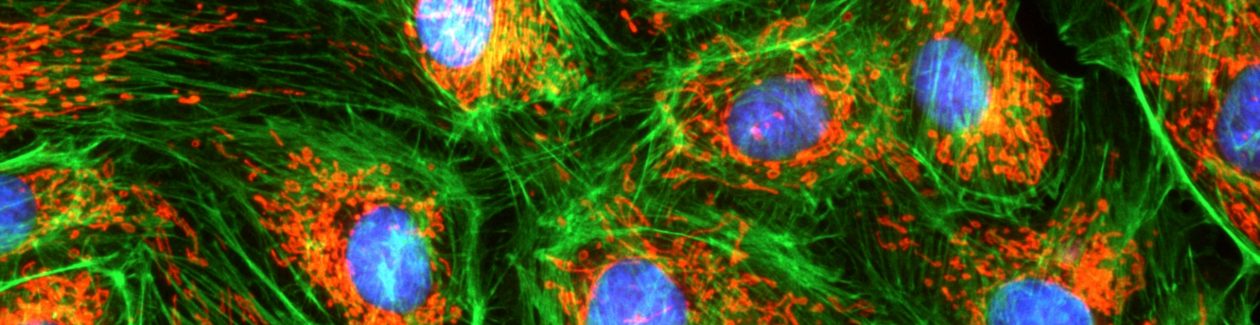
JNO-G(B)필터를 이용한 Sample Image 촬영

JNO-G(B)를 사용하여 보는 TRITC 형광 이미지
Filter Set : JNO-G(B)
Camera : AcquCAM 3G
C-mount adapter : 1x
Lens : UPLAPO20X
Sample : bovine pulmonary artery endothelial cell, BPAEC
대체 텍스트
Filter Set : JNO-G(B)
Camera : AcquCAM 5G
C-mount adapter : 1x
Lens : UPLAPO40X
Sample : bovine pulmonary artery endothelial cell, BPAEC
JNO-G(B)를 사용하여 보는 TRITC 형광 이미지
Filter Set : JNO-G(B)
Camera : AcquCAM 23GR2
C-mount adapter : 1x
Lens : UPLAPO20X
Sample : bovine pulmonary artery endothelial cell, BPAEC

JNO-G(B)필터에 적합한 형광색소


JNO-G(B) Spec

  • Typical Application(s) : Widefield Microscopy
  • Coating : Sputter/Hard Coated
  • Round excitation and emission filters mounted in anodized aluminum rings 2.3mm thick, up to 25mm in diameter (for standard sizes)
  • Rectangular dichroics up to 26x38mm, 1mm thick, to fit standard microscope manufacturer filter cubes (for standard sizes)

*** For filters larger than 25mm and dichroics larger than 26x38mm, please contact us for pricing.

JNO-B(B)

JNOPTIC Filter JNO-B(B)

Download Catalog _ JNO-B(B)

– Filter set for EGFP / FITC / Cy2 / AlexaFluor488
– Chroma Filter Model No. : 39002

JNO-B(B) Spectrum

FiltersType
AT480/30xEX
 AT505DCBS
AT535/40mEM

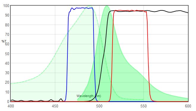
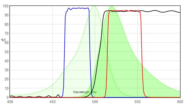
주요 형광 색소(Fluorochromes)와 스펙트럼 비교

* FITC VS JNO-B(B)

FITC VS JNO-B(B)

 * GFP VS JNO-B(B)

JNO-B(B) VS GFP spectrum 비교
JNO-B(B) VS GFP spectrum chart

 * AlexaFluor488 VS JNO-B(B)

JNO-B(B) VS AlexaFluor 488

JNO-B(B)필터를 이용한 Sample Image 촬영

Fluorescence Image by AcquCAM 23GR2
Sample : bovine pulmonary artery endothelial cell, BPAEC Filter Set : JNO-B(B) Camera : AcquCAM 23GR2 C-mount adapter : 1X Lens : PlanApo60xWLSM
Fluorescence Image by AcquCAM 3G
Sample : bovine pulmonary artery endothelial cell, BPAEC Filter Set : JNO-B(B) Camera : AcquCAM 3G C-mount adapter : 1X Lens : PlanApo60xWLSM
Fluorescence Image by AcquCAM 5G
Sample : bovine pulmonary artery endothelial cell, BPAEC Filter Set : JNO-B(B) Camera : AcquCAM 5G C-mount adapter : 1X Lens : UPLNAPO40X

JNO-B(B)필터에 적합한 형광색소


JNO-B(B) Spec

  • Typical Application(s) : Widefield Microscopy
  • Coating : Sputter/Hard Coated
  • Round excitation and emission filters mounted in anodized aluminum rings 2.3mm thick, up to 25mm in diameter (for standard sizes)
  • Rectangular dichroics up to 26x38mm, 1mm thick, to fit standard microscope manufacturer filter cubes (for standard sizes)

*** For filters larger than 25mm and dichroics larger than 26x38mm, please contact us for pricing. ( Tel. 02-3473-4188)

JNO-U(B)

Download Catalog _ JNO-U(B)

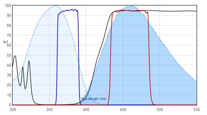
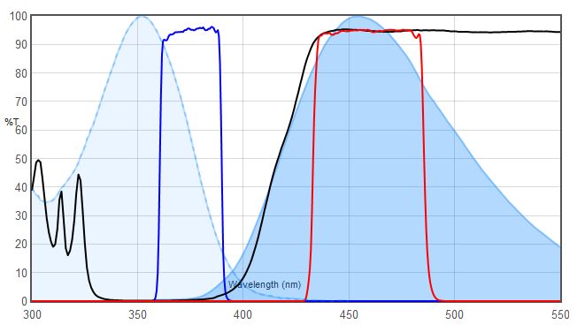
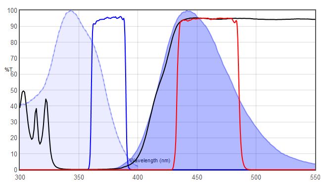
– Filter set for DAPI / Hoechst / AlexaFluor350
– Chroma Filter Model No. : 39000

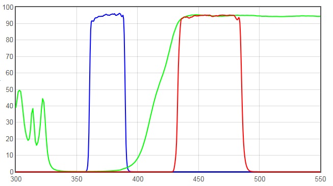
JNO-U(B) Spectrum

JNO-U(B)
Filters Type
 AT375/28x EX
AT415DC BS
 AT460/50m EM

주요 형광 색소(Fluorochromes)와 스펙트럼 비교

* DAPI VS JNO-U(B)

DAPI VS JNO-U(B)

* Hoechst VS JNO-U(B)

JNO-U(B) VS Hoechst

 * AlexaFluor350 VS JNO-U(B)

JNO-U(B) VS AF350

JNO-U(B)필터를 이용한 Sample Image 촬영

DAPI
Sample : bovine pulmonary artery endothelial cell, BPAEC
Camera : AcquCAM 23GR2, Filter Set : JNO-U(B)
Objective lens : UPLNAPO40X, C-mount adapter : 1X
AcquCAM 23GR2 fluorescence Image
DAPI, FITC, TRITC
Sample : bovine pulmonary artery endothelial cell, BPAEC
Camera : AcquCAM 23GR2, Filter Set : JNO-U(B),B(B),G(B)
Objective lens : UPLNAPO40X, C-mount adapter : 1X (OLYMPUS)
Above three images are merged and processed using JNO-ARM
DAPI, FITC, TRITC
Sample : bovine pulmonary artery endothelial cell, BPAEC
Camera : AcquCAM 3G, Filter Set : JNO-U(B),B(B),G(B)
Objective lens : UPLNAPO40X, C-mount adapter : 1X
Above three images are merged and processed using JNO-ARM

JNO-U(B) 필터에 적합한 형광색소

JNO-U(B) Spec

  • Typical Application(s) : Widefield Microscopy
  • Coating : Sputter/Hard Coated
  • Round excitation and emission filters mounted in anodized aluminum rings 2.3mm thick, up to 25mm in diameter (for standard sizes)
  • Rectangular dichroics up to 26x38mm, 1mm thick, to fit standard microscope manufacturer filter cubes (for standard sizes)

*** For filters larger than 25mm and dichroics larger than *** 26x38mm, please contact us for pricing.

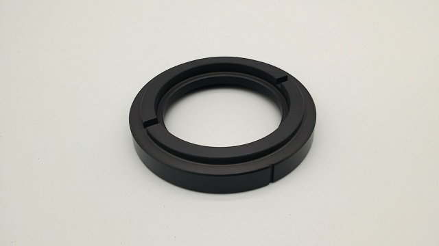
JNO-WICD-02

Filter Holder for BX50 WI

(OLYMPUS  – 32BP775)

45㎜ holder for 32㎜ filter
  •  45㎜ 필터용 위치에 32㎜ 필터를 사용할 수 있도록 만든  홀더
  • 여러개의 필터를 중첩하여 사용 가능함.
  • BX50-WI (Olympus)  광학 유닛 업그레이드시에 JNO-DICD-01과 같이 사용되어 지며,  32BP775 설치시에 필요한 홀더임.

Lecturer Profile

Profile of   Jin Jae-Hwan ( Instructor )

CEO of JNOpTIC co., ltd

June 25, 2021

Patent registration:
Observation method and observation device

Simultaneous observation
( Bright Field and Fluorescence microscopy
)

Fluorescene Microscopy (형광 현미경 검경법)과
Bright Field Microscopy (명시야 현미경 검경법)의
동시관찰을 위한 관찰 방법 및 관찰 장치

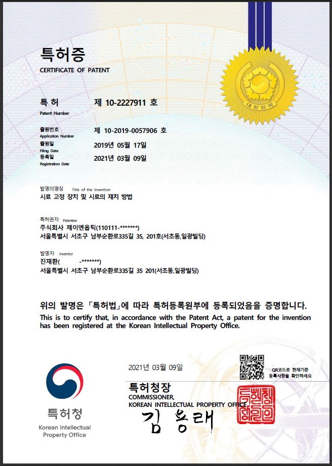
March 09, 2021

Patent registration:
sample fixing device and method of placing sample

Patent applied to section observation microscope
(JNO-FM-BX53-SET)

JNOPTIC Co.,ltd Partent
Sample holding device and method of placing sample
Sample fixing device for observing the cross section of the flexible packaging film and method of placing the sample (Patent)

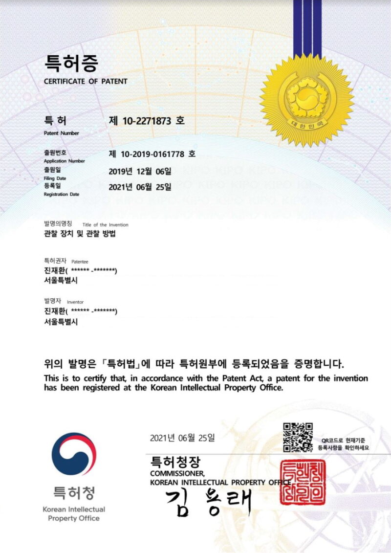
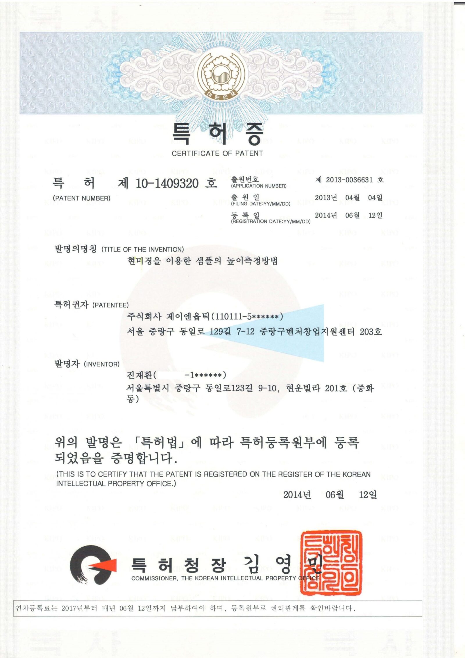
June 12, 2014

Patent registration:
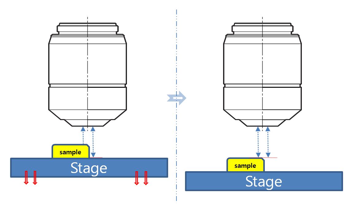
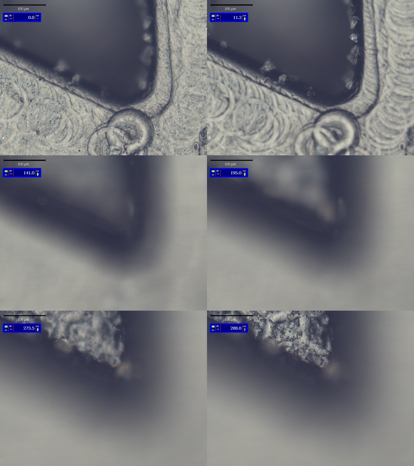
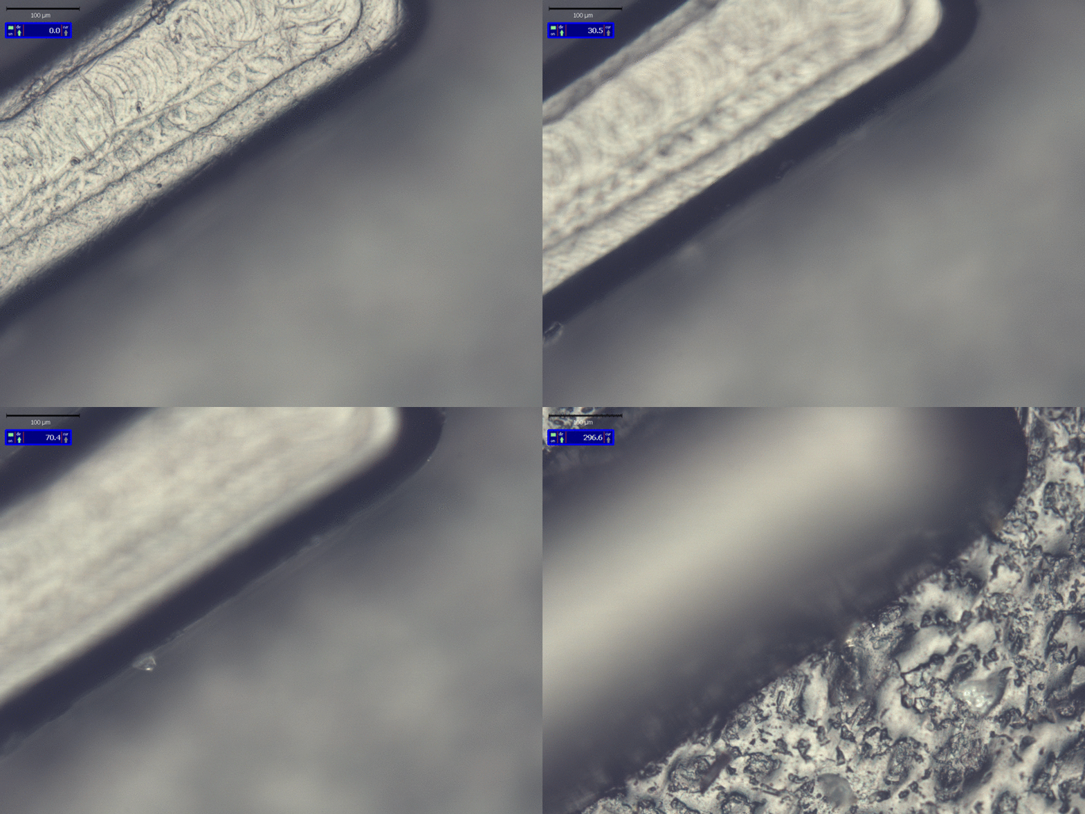
Method of measuring the height of a sample using a microscope

Certificate of Patent
Patent: Height measurement method of sample by microscope

현미경을 이용한 샘플의 높이측정방법

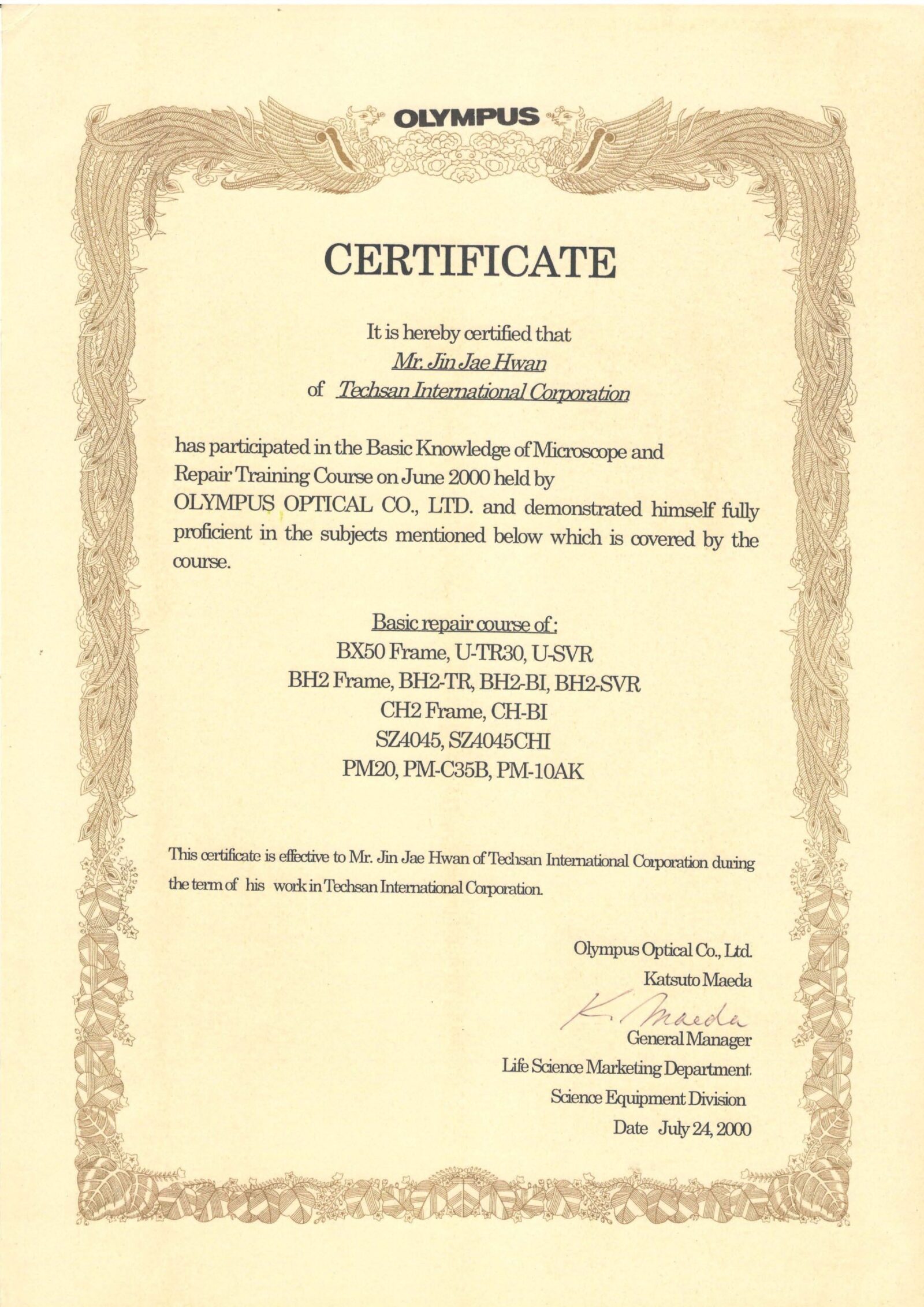
June 5th ~ June 15th, 2000 ( OLYMPUS Japan in Tokyo)

Basic Knowledge of Microscope and Repair Training Course


September 4, 2003 ( OLYMPUS Japan in Tokyo)

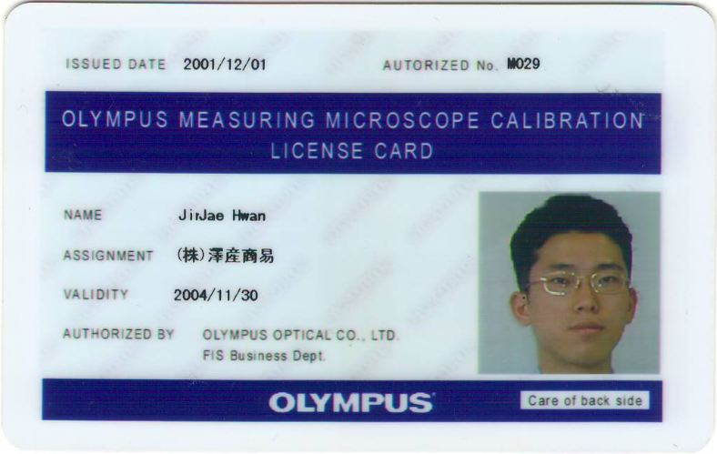
Measuring Microscope Calibration License

Main Model: STM6

License ID Number: M029


September 4, 2003 ( OLYMPUS Japan in Tokyo)

Biological Confocal Microscope Setup & Service Training

Main Model: FluoView300 & FluoView500


October 3, 2003 ( OLYMPUS Japan in Tokyo)

Inverted Microscope  & Research Stereo Microscope Service Training

Main Model: IX71 / SZX9


August 17th ~ August 20th, 2004 ( OLYMPUS Japan in Tokyo)

Confocal Microscope Setup & Service Training

Main Model:  FV1000


October 11 ~ October 13, 2004 ( OLYMPUS Japan in Tokyo)

Confocal Microscope Setup & Service Training

Main Model:  FV1000 M-COMB(Multi Combiner)


June 20, 2005 ~ June 23, 2005 ( OLYMPUS Japan in Tokyo)

Research System Microscope Service Training

 Main Model: AX70


August 1st-August 2nd, 2005 ( OLYMPUS Japan in Tokyo)

Polarizing Microscope Instruction Training

Main Model: BX51P


December 19, 2005 ( OLYMPUS Japan in Tokyo)

ZDC Training for Maintenance &  Basic Knowledge of Confocal Microscope


December 20 ~ 22, 2005 ( OLYMPUS Japan in Tokyo)

Brushup Course to bring out the power of a microscope (GA Academy)


August 2nd ~ August 4th, 2006 ( OLYMPUS Japan in Tokyo)

Confocal Laser Scanning Microscope for Industrial Market

D class License(for ols3000)

Main Model:  OLS 3000(Lext)


October 11 ~ October 15, 2006 ( OLYMPUS Japan in Tokyo)

Confocal Laser Scanning Microscope Training for Bio Maket

Maintenance Service Training

Main Mode :  M-COMB(Multi Combiner for FV1000)


December 9, 2006

IX Repair training


May 07 ~ May 11, 2007 ( OLYMPUS Japan in Tokyo)

Modify Training for Zero-Drift Compensation Unit of IX81

IX2-Cusominsing Training (Optic Port Modify)

Main Model: IX71 & IX81-ZDC


September 3rd ~ September 08th, 2007 ( OLYMPUS Japan in Tokyo)

Leicense D class Trainging

Main Model: OLYMPUS Bio Confocal Microscope  FV1000


April 08 ~ April 10, 2008 ( OLYMPUS Japan in INA_Nagano )

IV100 Setup Training

(in vivo fluorescence molecular imaging systems)


July 22 ~ July 23, 2008  ( Narishige Group in Tokyo,  Japan )

 Narishige Maintenance Training


July 24th ~ July 28th, 2008 ( OLYMPUS Japan in Tokyo)

Research Inverted Microscope  maintenance Training

Main Model: IX81


May 18 ~ May 22, 2009 ( OLYMPUS Japan in Tokyo)

OLYMPUS Bio Confocal Microscope  License Training

 License Grade: FV1000 C class


June 23 ~ June 26, 2009 ( OLYMPUS Japan in Tokyo)

Confocal  Laser Scanning  Microscope  License Training

License Grade: FV10i  C class


October 19 ~ October 23, 2009 ( OLYMPUS Japan in Tokyo )

Multi Photon Laser Scanning MICROSCOPE Setup Training

Main Model: MPE C class


August 3rd ~ August 8th, 2010 ( OLYMPUS Japan in Tokyo )

Modify Training for Zero-Drift Compensation Unit of IX81

Main Model: IX81-ZDC2

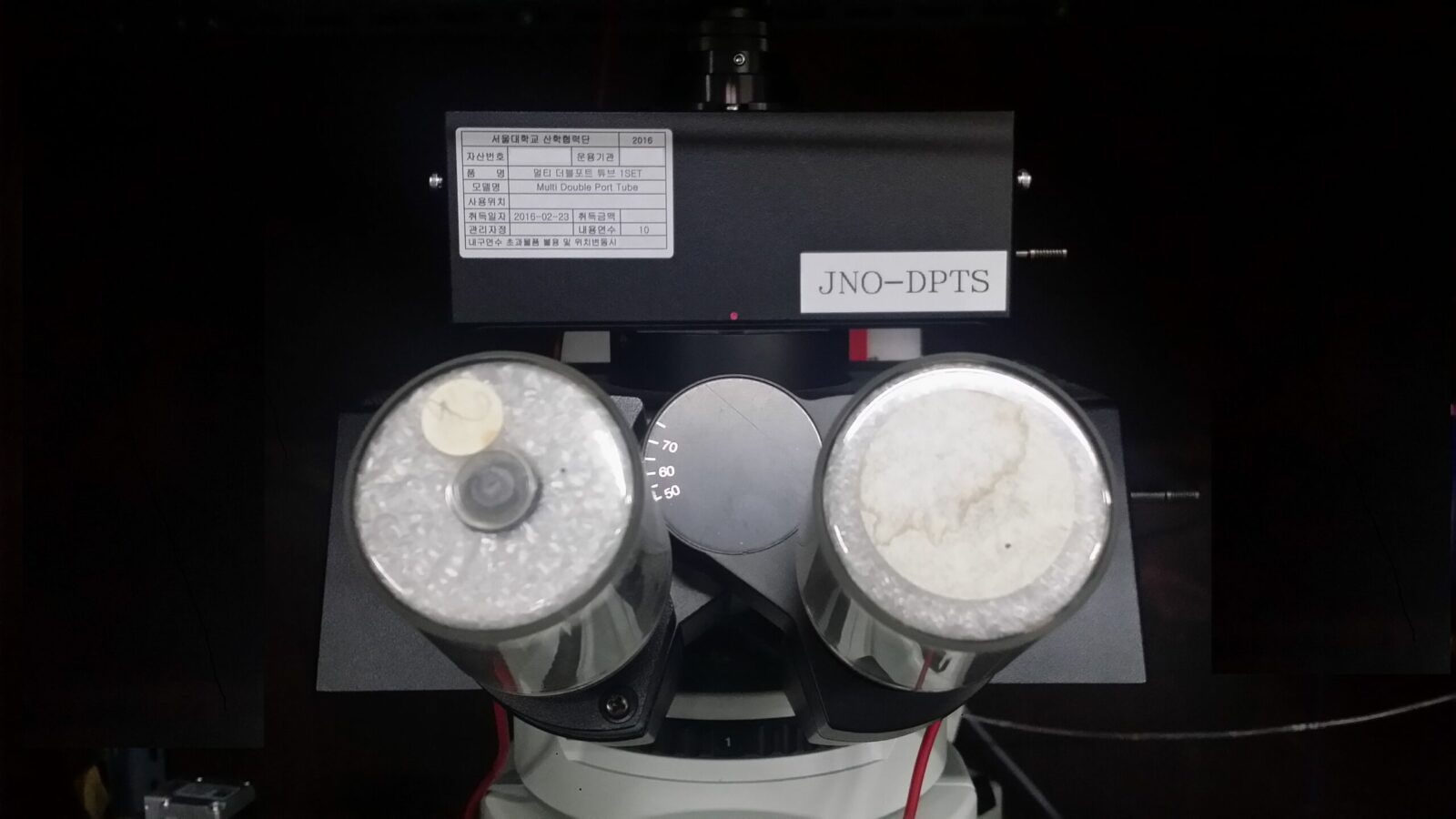
JNO-DPTS

JNO-DPTS (Down load Catalog JNO-DPTS)

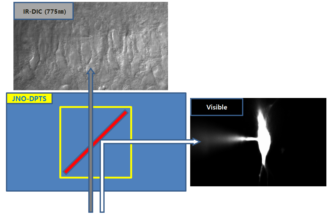
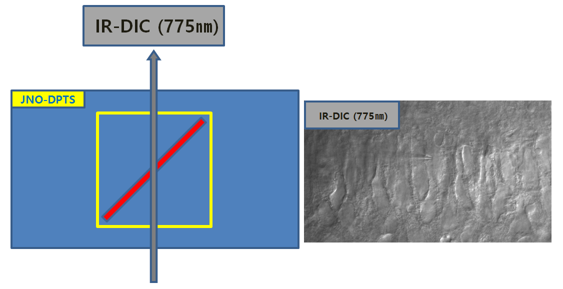
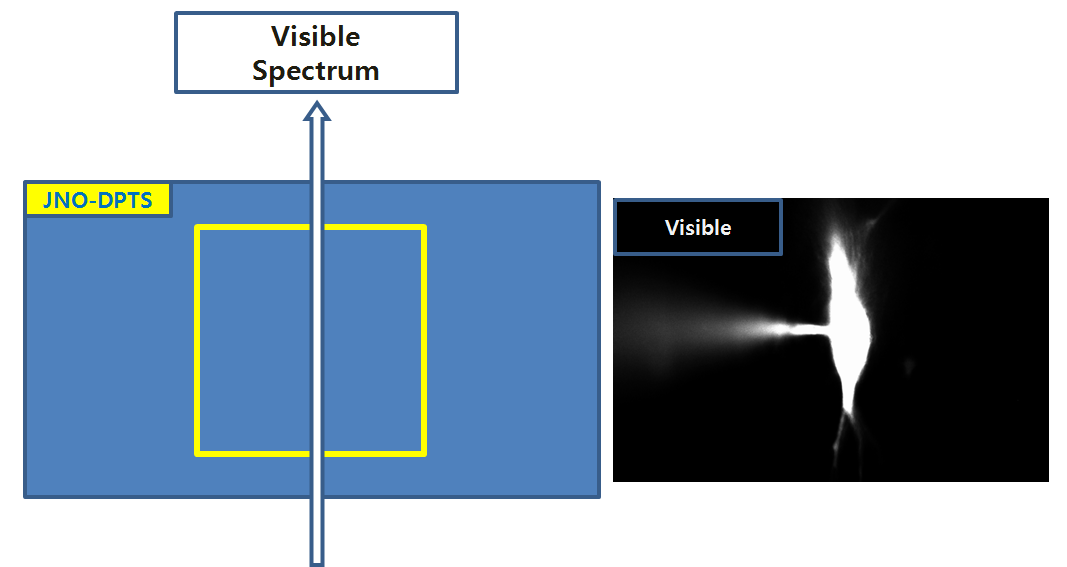
The Unit to observe fluorescence image and IR DIC image at the same time with Multi-Dual Port tube of BX51-WI
(Made by JNOpTIC co.,ltd )

JNOPTIC Multi-Dual Port System
  1. Simultaneously Observation

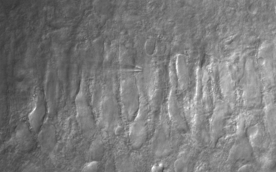
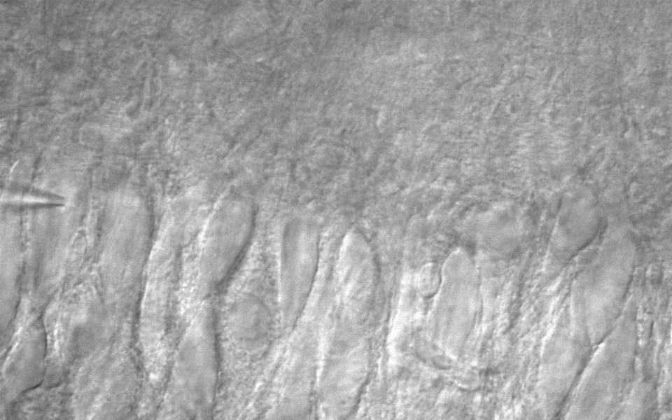
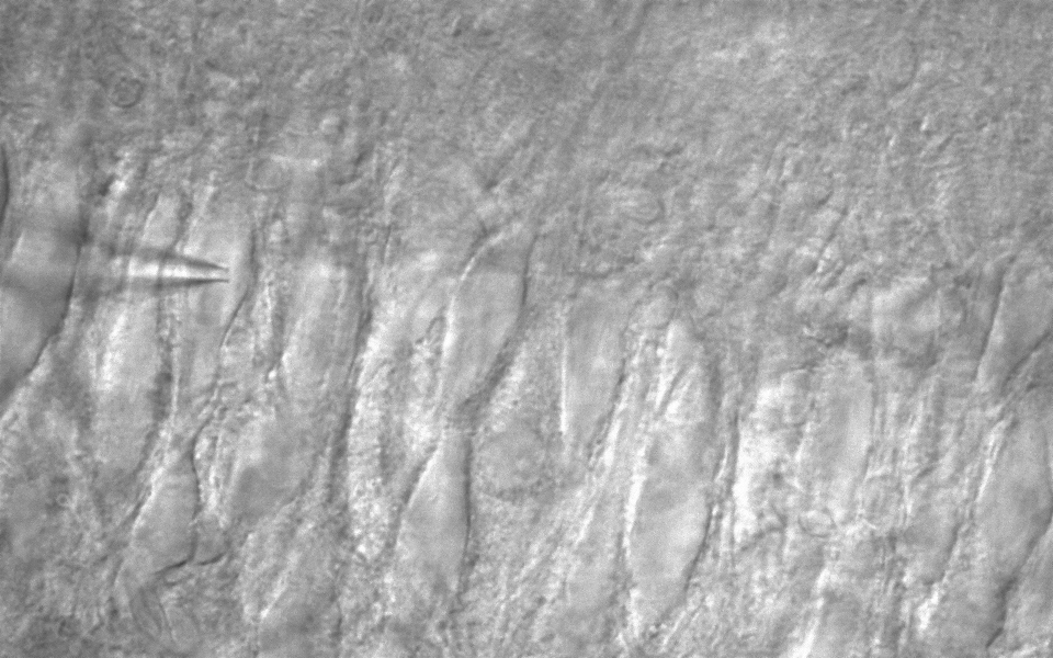
2-1. One Way((IR-DIC 775nm)

2-2. One Way((Visible Spectrum)

Simultaneously observation
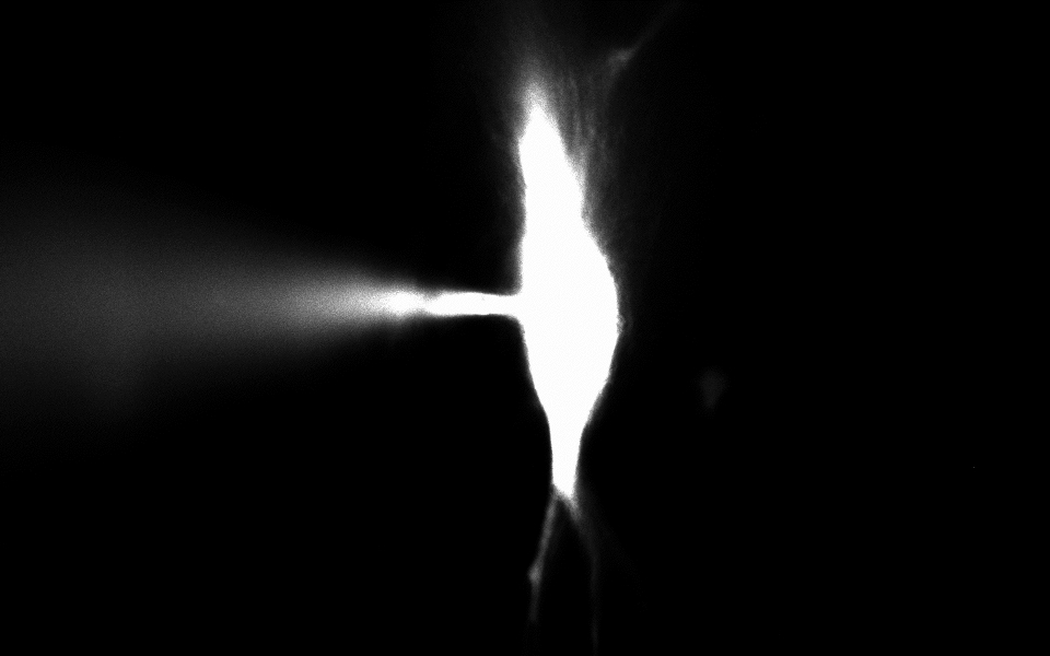
– CA3 pyramidal neuron, IR-DIC & Alexa Fluor 488 (50 uM)

20160323_1(FullF6)_2 for wp

20160323_1(1) for wp
CA3 pyramidal neuron Alexa Fluor 488, IR-DIC (Simultaneous observations)
Figure courtesy by Dr. Sooyun Kim, Seoul National University
20151209_2_stim1(CA3 pyramidal neuron) for wp
CA3 pyramidal neuron AcquCAM 23S & JNO-RNB filter with BX51WI
(Download Catalog AcquCAM 23S)
20151209_2(CA3 pyramidal neuron) for wp
CA3 pyramidal neuron AcquCAM 23S & JNO-RNB filter with BX51WI
(Download Catalog AcquCAM 23S)

JNO-ARM

Advance Realtime Monitor. Easy-to-Use, Image Enhancement, etc

1. Information

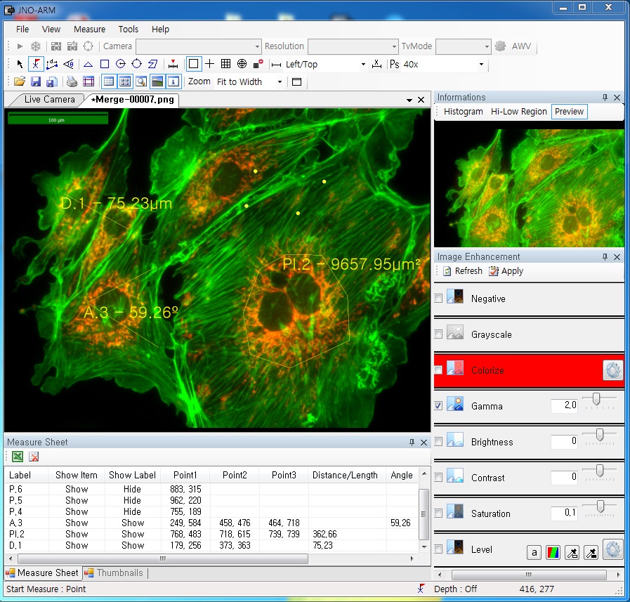
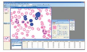
ARM is image analysis software for JNOPTIC AcquCAM cameras. This S/W is interchangeable with all WDM cameras regardless of camera brand and model and even more the most strength point is simple and easy use of length, area, angle, etc.ARM01

2. Simple measurement tools

toolbar

  • Measurement Tools: Count, Distance, Angle, Area, etc.
  • Guide Lines: Cross Line, Rectangular Lattice, Circular Grid
  • Scale Panel on Live/Image Screen
  • Available measurement on LIVE image and saved image mode

3-1. Specialization for observation of fluorescence images

Improvement Function about Fluorescence Microscopy Image. (Used function: Auto Level, Low level, Pseudo Color, Merge Image)
ARM02
 Bottom on the left is merged image without improvement of image. And the bottom on the right is merged image after improvement of image. These images are shot by JNOPTIC AcquCAM Pro/G3 camera

3-2-1 Effect of high-contrast image with simple operation (Monochrome)

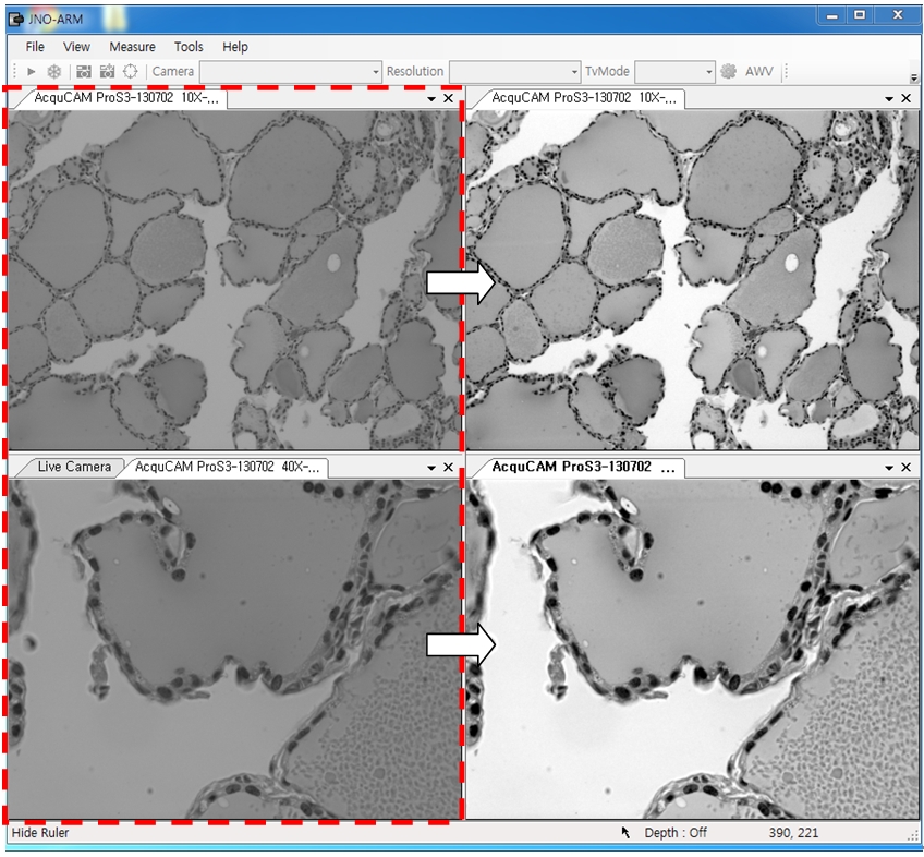
Improvement Function about Microscopy Image Monochrome. (Used function: Auto Level)arm04Effective improvement of image with simple operation. This image is shot by JNOPTIC Pro/S3 camera

3-2-2 Effect of high-contrast image with simple operation (Color)

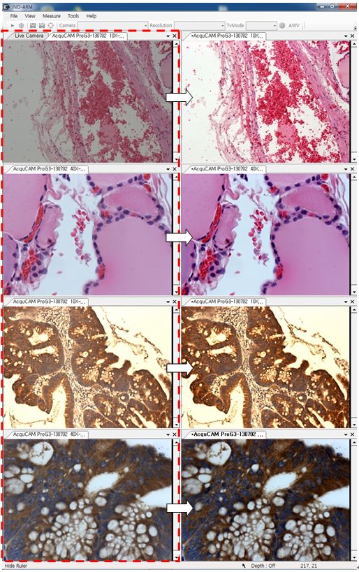
Improvement Function about Microscopy Image. (Used function: Auto Level)arm05Effective improvement of image with simple operation. These images are shot by JNOPTIC AcquCAM Pro/G3 camera

3-3-1 Live Pseudo Color (for color camera)

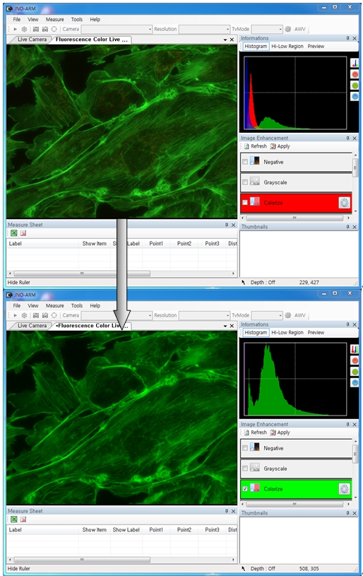
When you get fluorescence images using color camera, if the unwanted channel was shown because of cross talk, you could remove this channel to take advantage of function of Live Pseudo.
Improvement Function about Fluorescence Microscopy Live Camera Image. (Used function: Live Pseudo Color)
arm06
 The extraction of wanted color information from color image (Remove BR on RGB source) These images are shot by JNOPTIC AcquCAM Pro/G3 camera

3-3-2 Live Pseudo Color (for Monochrome camera)

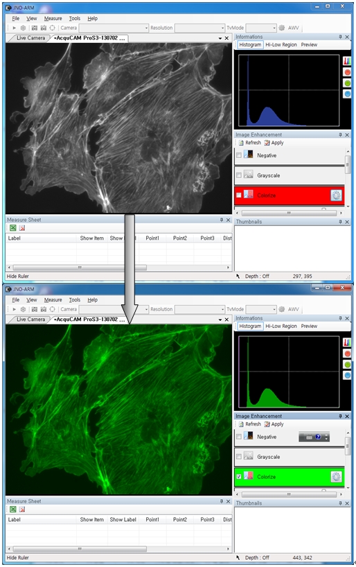
When you get fluorescence images using monochrome camera, you can raise efficiency of acquisition for fluorescence images to add similar false color to the color to be shown on the eyepiece in real time.
Improvement Function about Fluorescence Microscopy Live Monochrome Camera Image. (Used function: Live Pseudo Color)
arm08
 Improvement of usage environment for monochrome camera to add false color on the image. These images are shot by JNOPTIC AcquCAM Pro/S3 camera

4. JNO-MHU(Option Unit)

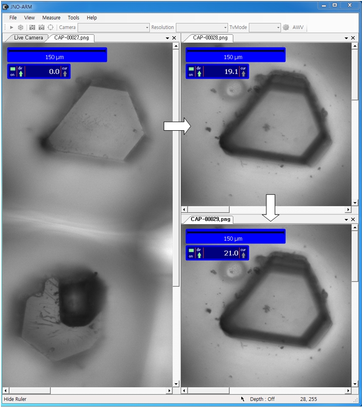
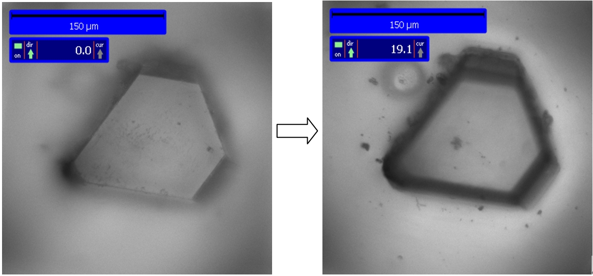
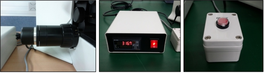
Available measuring height with additional unit, JNO-MHU
※ JNO-MHU is sold separately as optional unit.
jno-mhu1
  jno-mhu2
 Left: Focus on top of sample (Z-axis reset), Right: Focus on bottom of sample (Z-axis height measurement)
jno-mhu3
 < Measuring condition : over 20x objective, temp 20°C >
Responding Model Measurement value unit Measurable height Error of measuring 1000㎛ Recommended measuring height
CX 41 0.2 ㎛ -9.9 ~ 29.9㎜ Below ±20㎛ Below ± 2000㎛
CKX 41 0.2 ㎛ -9.9 ~ 29.9㎜ Below ±20㎛ Below ± 2000㎛
BX – FM 0.2 ㎛ -9.9 ~ 29.9㎜ Below ±20㎛ Below ± 2000㎛
BX 51/53 0.1 ㎛ -9.9 ~ 29.9㎜ Below ±10㎛ Below ± 1000㎛
MX 51 0.1 ㎛ -9.9 ~ 29.9㎜ Below ±10㎛ Below ± 1000㎛
MX 61L/61 0.1 ㎛ -9.9 ~ 29.9㎜ Below ±10㎛ Below ± 1000㎛

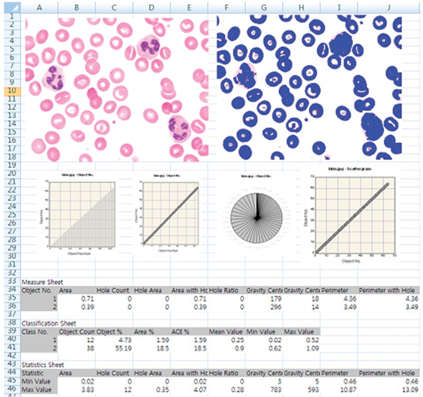
5. Reporting to Excel

excel


OpTIC Eye (Auto)

JNOPTIC Image Analysis Software OpTIC Eye Series

Catalog_OpTIC Eye

  1. OpTIC Eye (Auto)

기본측정+보고서+오토카운팅(입도분석용)
– 셀이미지 자동측정(5단계 easy측정)
: 영상분할, 측정항목설정, 측정, 분류, 성적서기능
– 면적, 면적비, 반경, 직경등 15개 항목 자동측정

Auto Counting

영상을 자동 분석하는 기능입니다. 영상을 개체들(Objects)과  배경(Background)으로 분할(Segmentation)함 으로써 측정할 영역을 한번에 검출해내고, 면적 등을 자동측정 합니다. Auto Count는 위의 차례대로 측정이 이루어 집니다.

  • 영상분할(Segmentation)
  • 측정항목설정(Define Measure)
  • 측정(Measure)
  • 분류(Classification)
  • 성적서(Report)


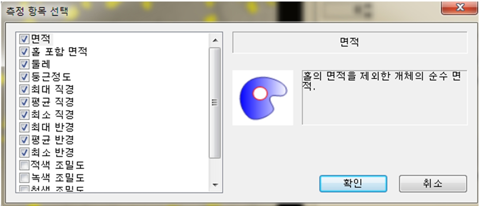
측정 항목 설정(Define Measure)

1

  • 원하는 측정 항목 설정
  • 측정항목별 그림에 의한 상세설명
  • 구간 설정: 상한, 하한 적용가능


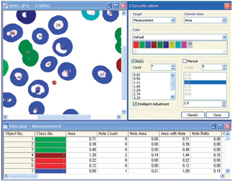
측정결과를 분석/분류 (Classification)

23

View Measurement를 통한 실시간 측정 항목 display 측정

크기별로 그룹화(Classification), 그룹별 색상화 가능


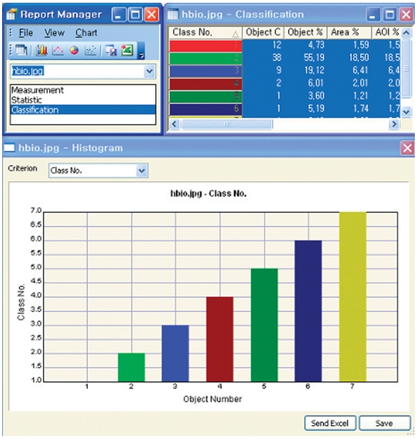
Report

4

측정결과를 4가지 형태의 Chart로 표시: Histogram, Line graph, Pie, Scatter-gram Excel 전송 가능