Spectrum Chart

Spectrum Chart
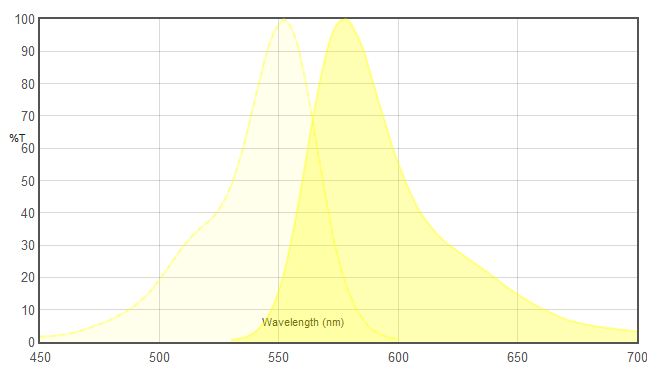
| Excitation (㎚) | Emission (㎚) |
| 555 | 580 |
| Filter Model No. | Excitation filter (㎚) | Dichroic mirror (㎚) | Emission filter (㎚) | Remarks |
| JNO-G(B) | 540/25 (527.5~552.5) |
565 (LP) | 605/55 (577.5~632.5) |
Narrow Excitation band, Bandpass Emission |
| 49004 | 545/25 (532.5~557.5) |
565 (LP) | 605/70 570~640) |
Narrow Excitation band, Bandpass Emission |
| 49305 | 546/22 (535~557) |
565 (LP) | 590/33 (573.5~606.5) |
Narrow Excitation band, Bandpass Emission |
| Filter Model No. | Excitation filter (㎚) | Dichroic mirror (㎚) | Emission filter (㎚) | Remarks |
| 49908 | 532/10 (527~537) |
532 (LP) | 590/50 (565~615) |
For use with laser between 529~535㎚
Very Narrow Excitation band, |
| 49909 | 561/10 (556~566) |
561 (LP) | 600/50 (575~625) |
For use with laser between 557~563㎚
Narrow Excitation band, |
| Excitation (㎚) | Emission (㎚) |
| 490 | 525 |
| Filter Model No. | Excitation filter (㎚) | Dichroic mirror (㎚) | Emission filter (㎚) | Remarks |
| JNO-B(B) | 480/30 (465~495) |
505 (LP) | 535/40 (515~555) |
Medium Excitation band, Bandpass Emission |
| 49002 | 470/40 (450~490) |
495 (LP) | 525/50 (500~550) |
Wide Excitation band, Bandpass Emission |
| 49011 | 480/40 (460~500) |
510 (LP) | 535/50 (510~560) |
Wide Excitation band, Bandpass Emission |
| Filter Model No. | Excitation filter (㎚) | Dichroic mirror (㎚) | Emission filter (㎚) | Remarks |
| 49904 | 488/10 (483~493) |
488 (LP) | 525/50 (500~550) |
For use with laser between 485~491㎚
Very Narrow Excitation band, |
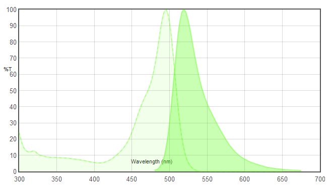
Fluorescein isothiocyanate (FITC) is a derivative of fluorescein used in wide-ranging applications including flow cytometry. FITC is the original fluoresce in molecule functionalized with an isothiocyanate reactive group (-N=C=S), replacing a hydrogen atom on the bottom ring of the structure. This derivative is reactive towards nucleophiles including amine and sulfhydryl groups on proteins.
A succinimidyl-ester functional group attached to the fluorescein core, creating “NHS-fluorescein”, forms another common amine reactive derivative that has much greater specificity toward primary amines in the presence of other nucleophiles.
FITC has excitation and emission spectrum peak wavelengths of approximately 495 ㎚ /519 ㎚. Like most fluorochromes, it is prone to photobleaching. Because of the problem with photobleaching, derivatives of fluorescein such as Alexa 488 and DyLight 488 have been tailored for various chemical and biological applications where greater photostability, higher fluorescence intensity, or different attachment groups are needed.
| Excitation (㎚) | Emission (㎚) |
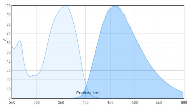
| 359 | 461 |
| Filter Model No. | Excitation filter (㎚) | Dichroic mirror (㎚) | Emission filter (㎚) | Remarks |
| JNO-U(B) | 375/28 (361-389) | 415 (LP) | 460/50 (435-485) | Narrow Excitation band, Bandpass Emission |
| 49000 | 350/50 (325-375) | 400 (LP) | 460/50 (435-485) | Wide Excitation band, Bandpass Emission |
| 49028 | 395/25 (382.5-407.5) | 425 (LP) | 460/50 (435-485) | For 395㎚ light sources Narrow Excitation band, Bandpass Emission |
DAPI (4′,6-diamidino-2-phenylindole) is a fluorescent stain that binds strongly to A-T rich regions in DNA. It is used extensively in fluorescence microscopy. As DAPI can pass through an intact cell membrane, it can be used to stain both live and fixed cells, though it passes through the membrane less efficiently in live cells and therefore the effectiveness of the stain is lower.
DAPI (4 ‘, 6-diamidino-2-phenylindole)는 DNA의 A-T가 풍부한 부위에 강하게 결합하는 형광 염료입니다. 그것은 형광 현미경에서 광범위하게 사용됩니다. DAPI는 손상되지 않은 세포막을 통과 할 수 있기 때문에 살아있는 세포에서 덜 효율적으로 막을 통과하기 때문에 살아있는 세포와 고정 된 세포를 모두 염색하는 데 사용할 수 있으므로 얼룩 효과는 낮습니다.
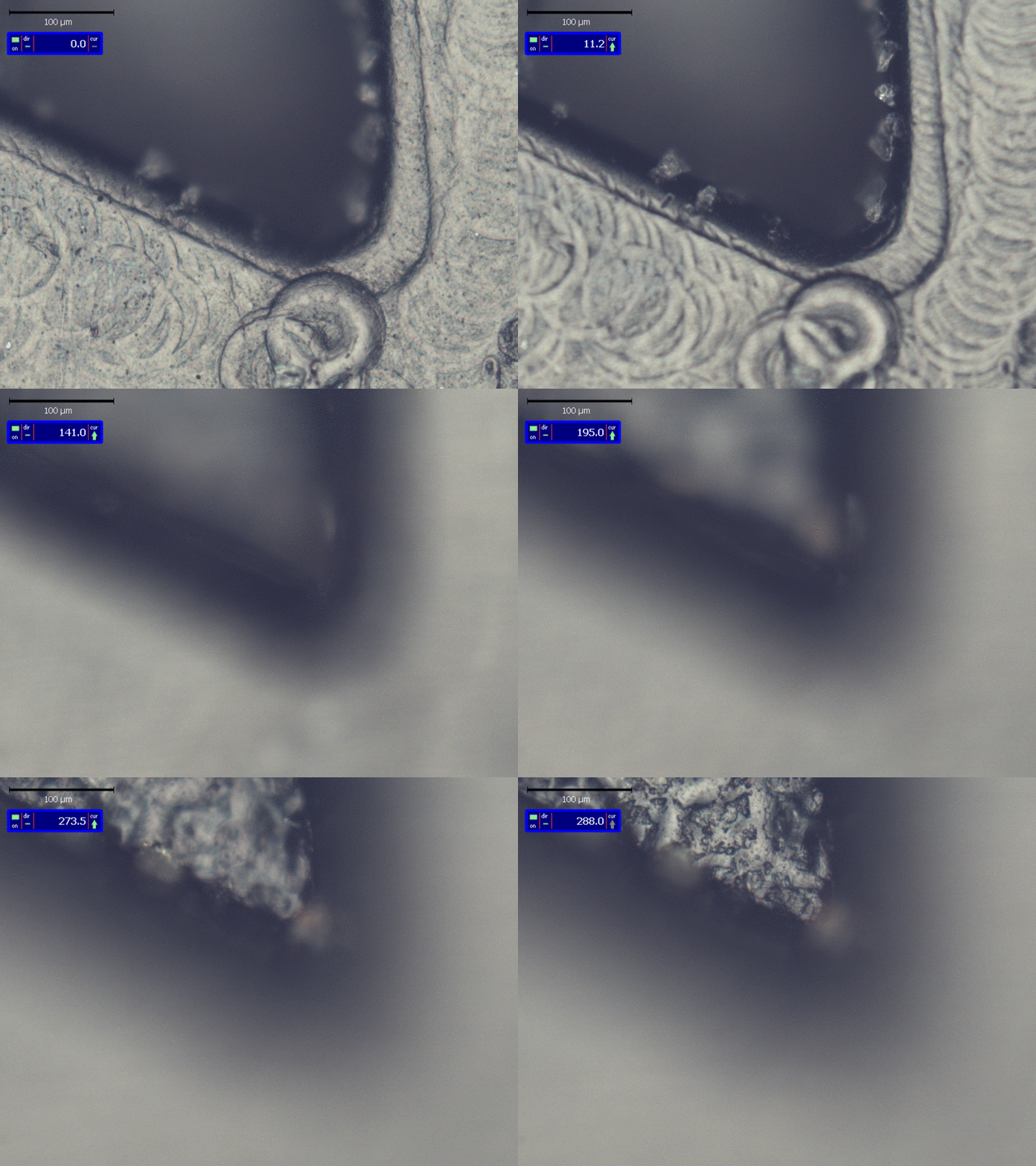
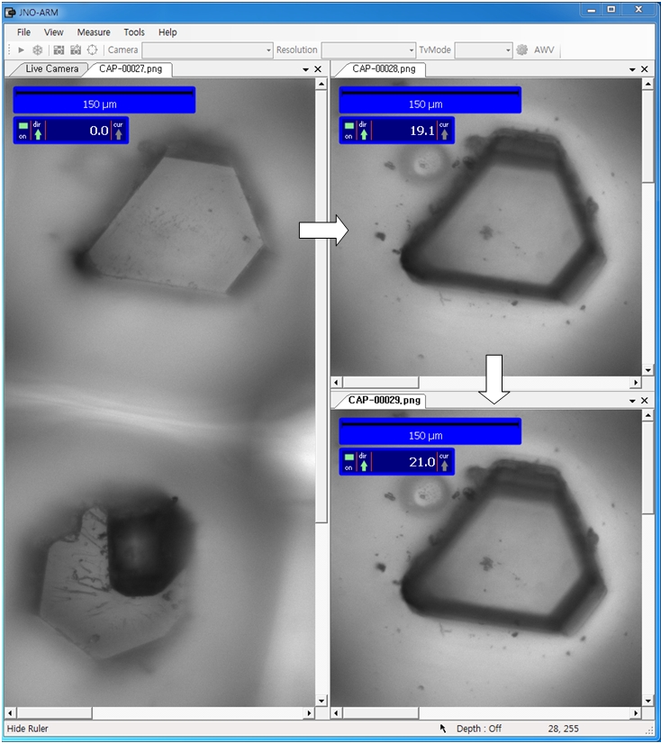
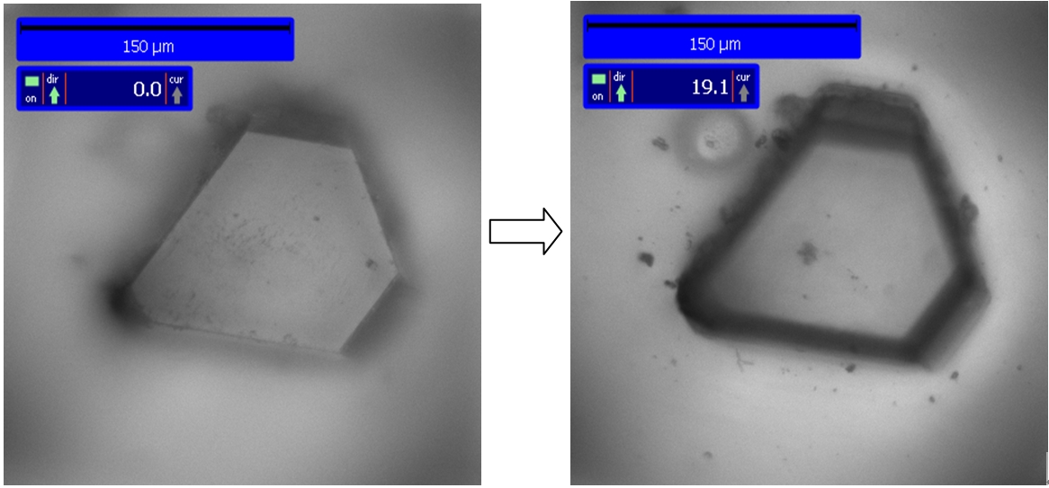
Right Image : Measurement of Z axis(height value) Z= 288㎛
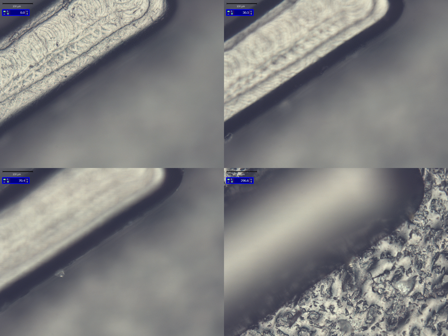
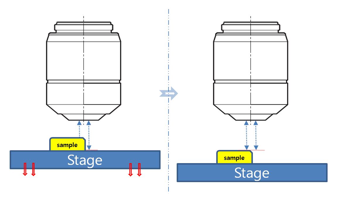
Heigh Measurement for Microscope
by JNO-MHU & JNO-ARM
| Certificate of Patent | Go to Main Page |
If you are interested in stock we have, please feel free to contact us.
E-mail : jhjin@jnoptic.com / Tel : 82-2-3473-4188
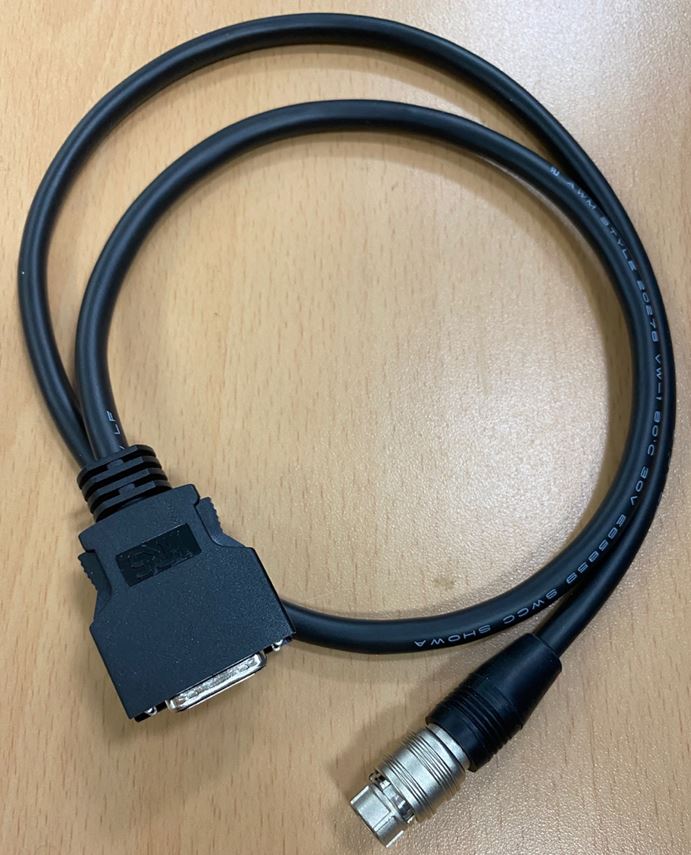
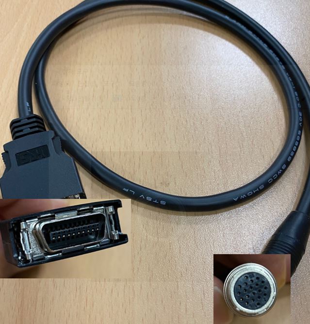
STM6 X-Y data cable
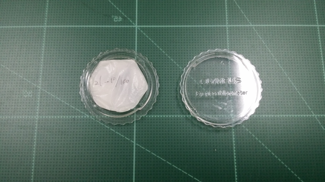
26OCM10/100
30.5S-G533
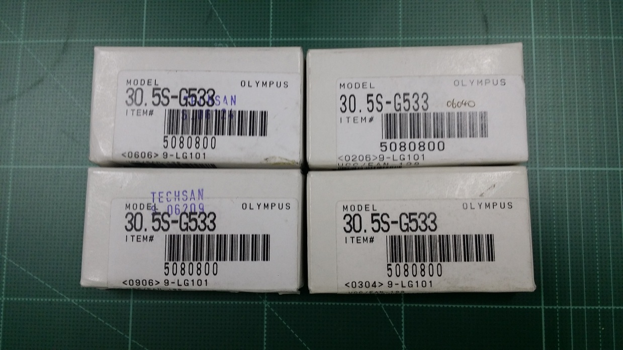
A 40 PL

AA1276 (SOLD OUT)
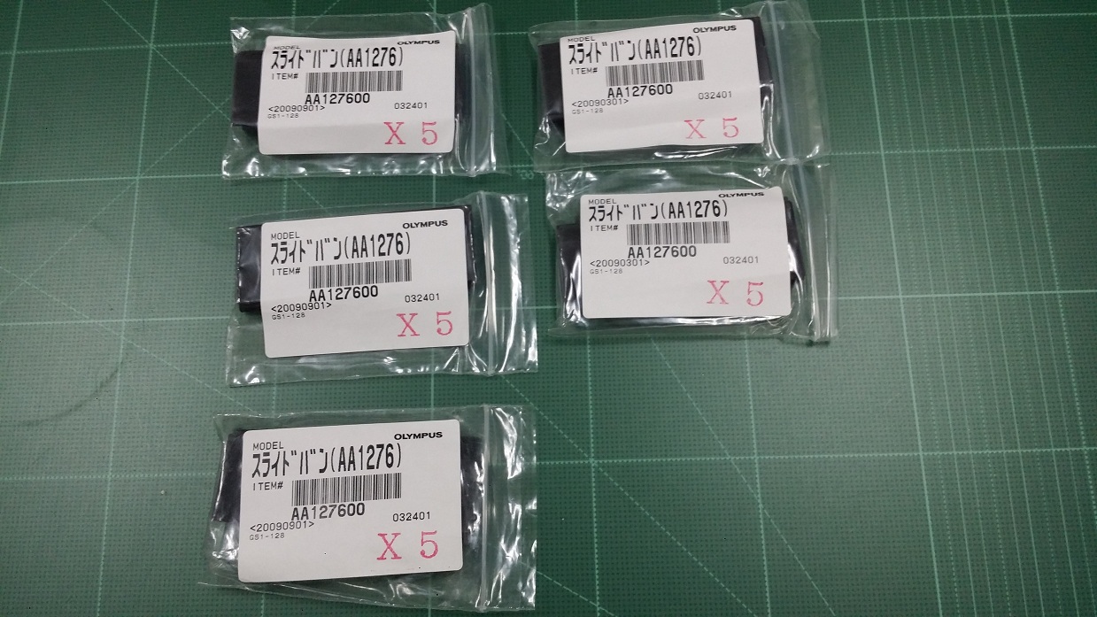
ADOBE PHOTOSHOP ELEMENTS 2.0

AL-1824
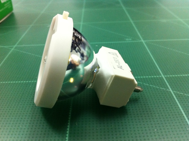
U-TV0.5X (SOLD OUT)
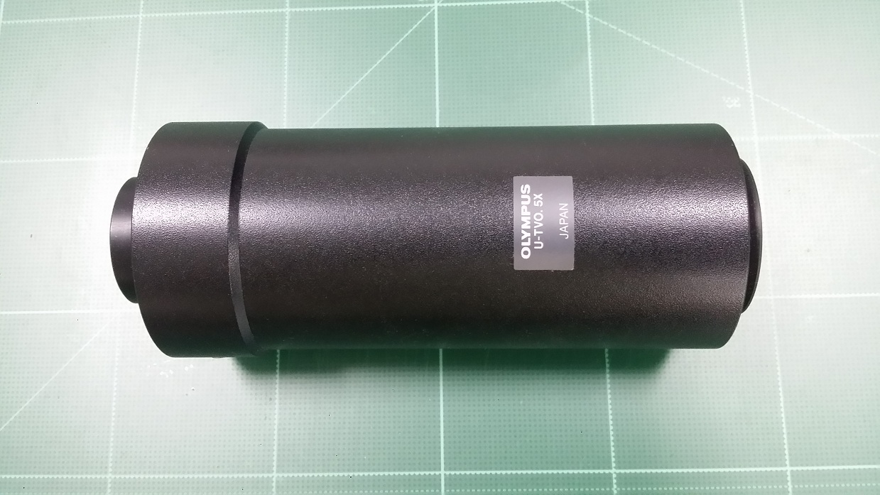
DP70-IFAD

FLM6
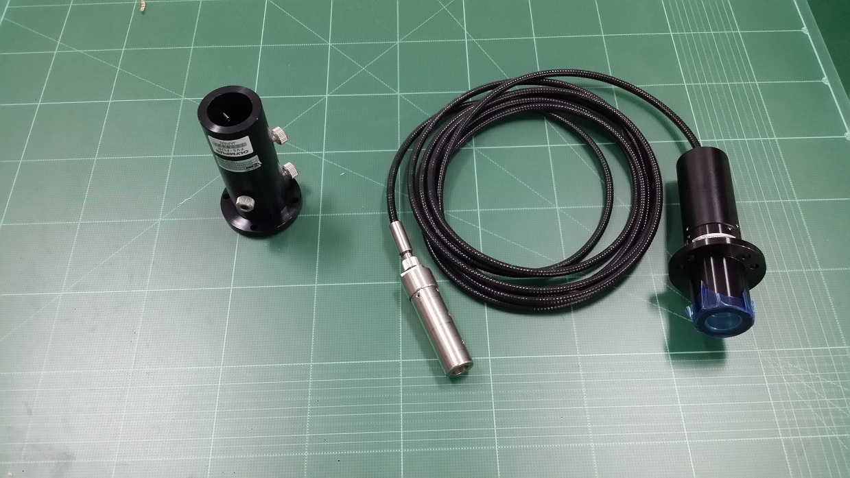
FV5-COL
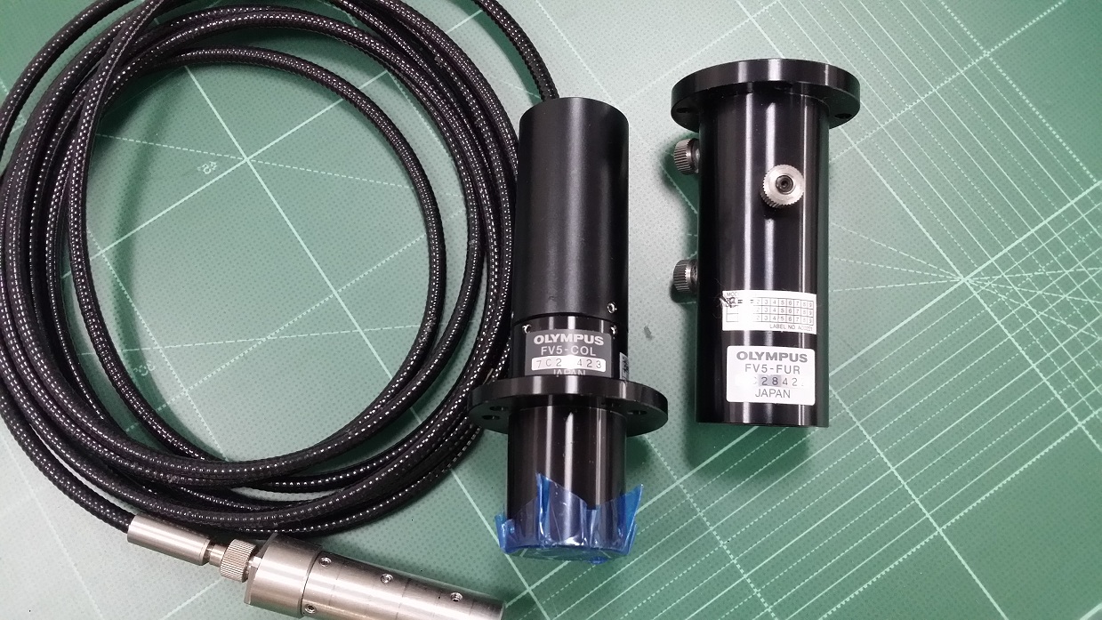
FV5-FUR
IX-DP10

IX-DP40

IX-DPA20

IX2-ARCEVA

IX2-DIC10
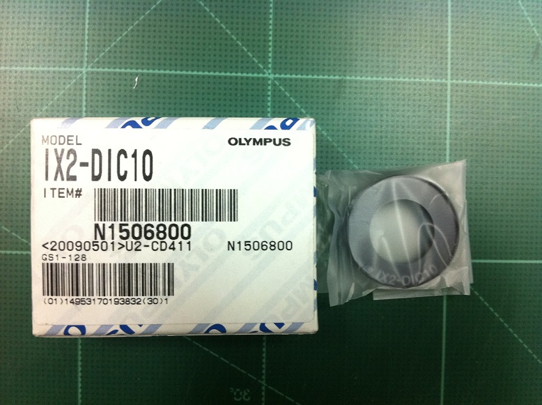
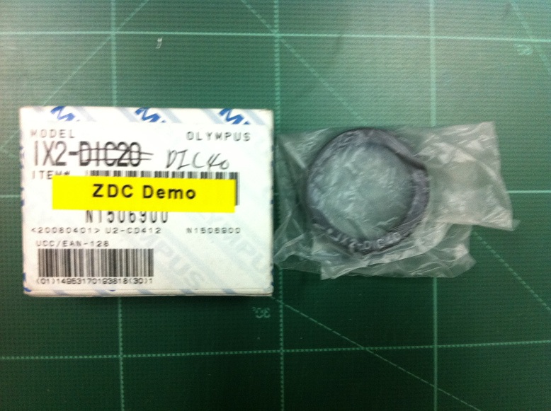
IX2-DIC40
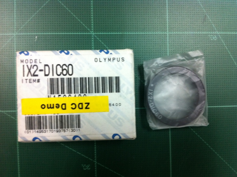
IX2-DIC60
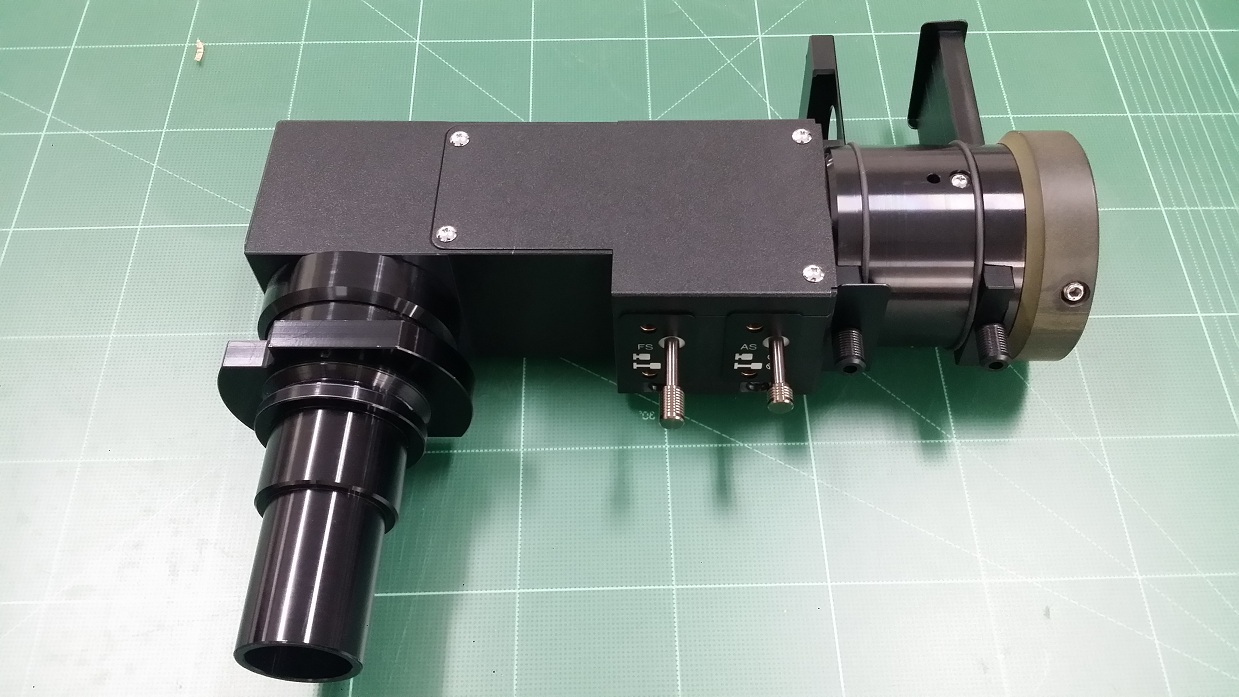
IX2-RFAL (SOLD OUT)
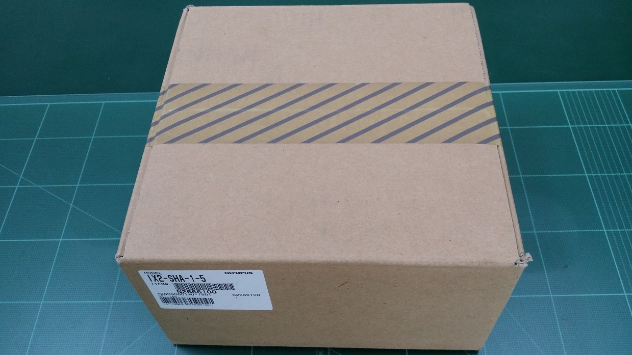
IX2-SHA-1-5
IX-SCL
IX-SUSP

LG-DI

LN22-P-13N1B

MM6-RHS250-2 (SOLD OUT)

PCDA40XPL-6

PL20
QUICKCAM FAST

RX-XYST

SZ2-LGSI
SZH-CLJ

SZX-APT (SOLD OUT)
SZX-AS (SOLD OUT)
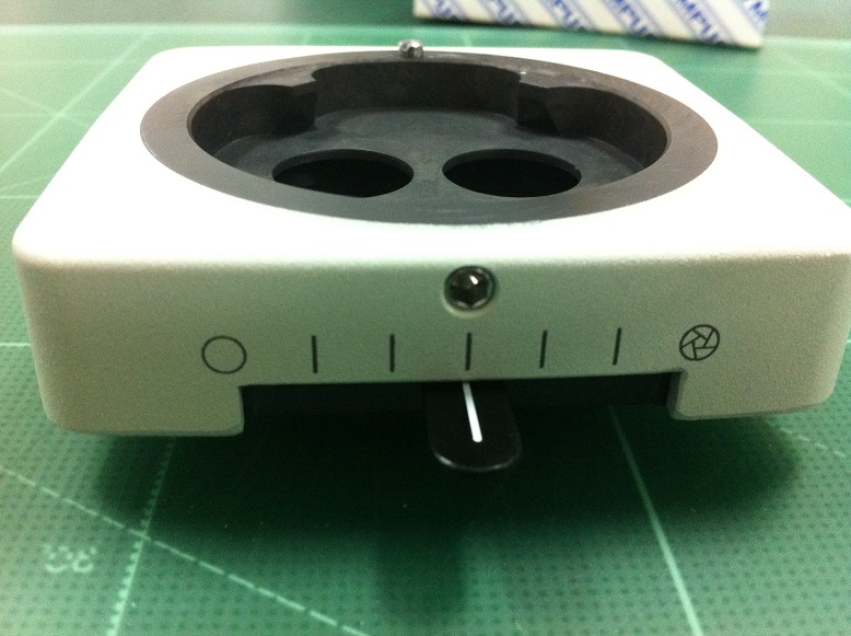
SZX-STAD1 (SOLD OUT)
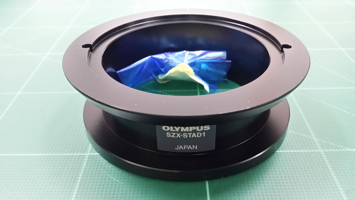
SZX-STL (SOLD OUT)
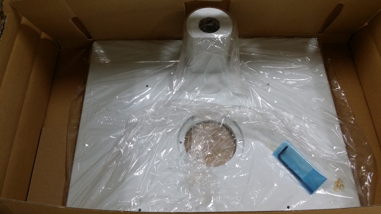
U-ACAD4515-1-5
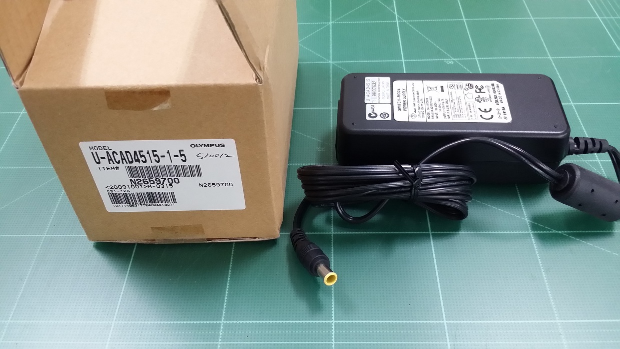
U-APT (Without Power cord)_(SOLD OUT)

U-DO3 (SOLD OUT)
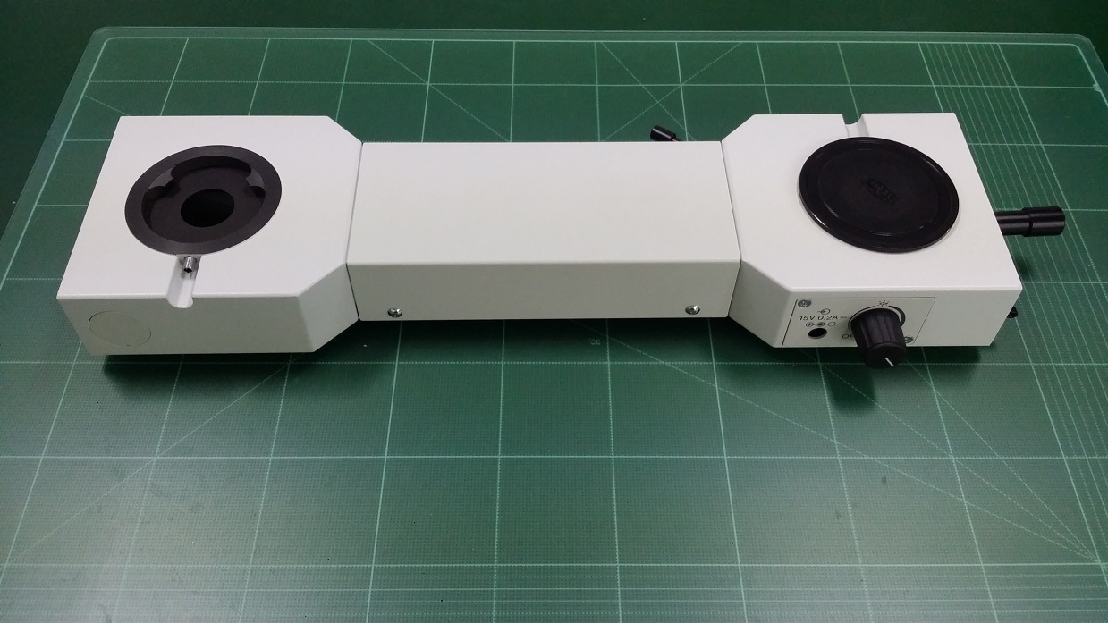
U-HSTR2
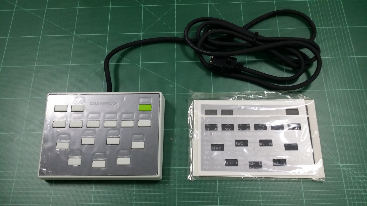
U-OSM
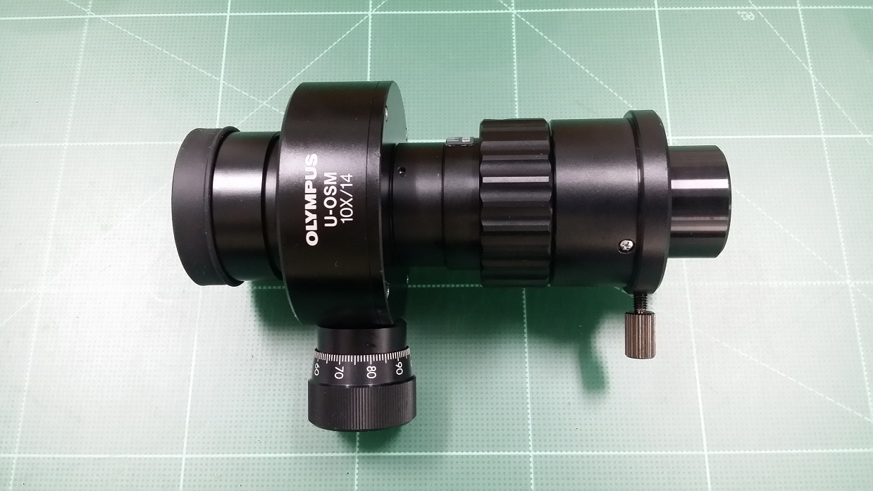
WI-FSH

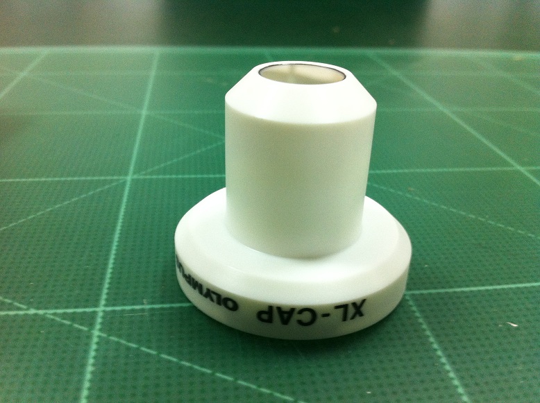
XL-CAP
PLAPO60XWLSM (SOLD OUT)
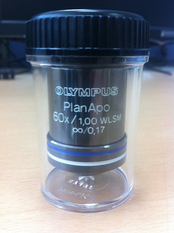
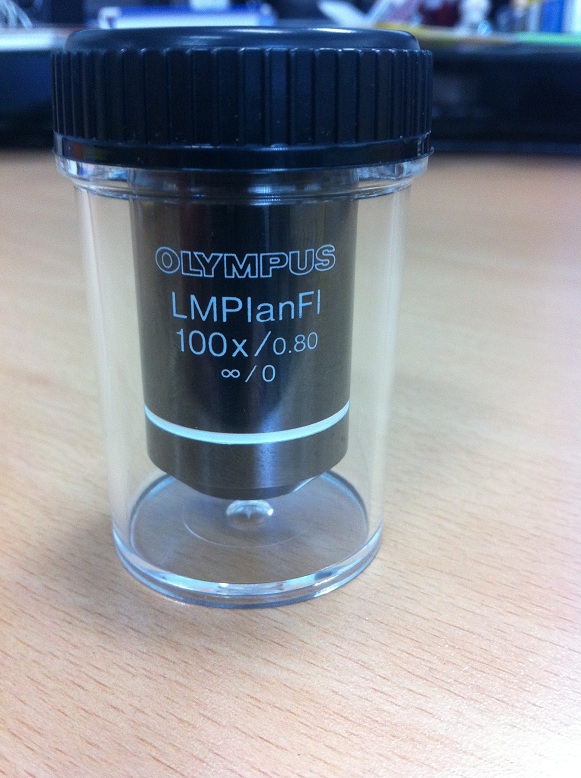
LMPLFL100X
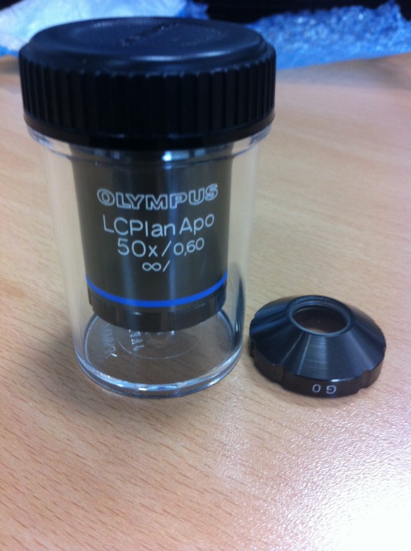
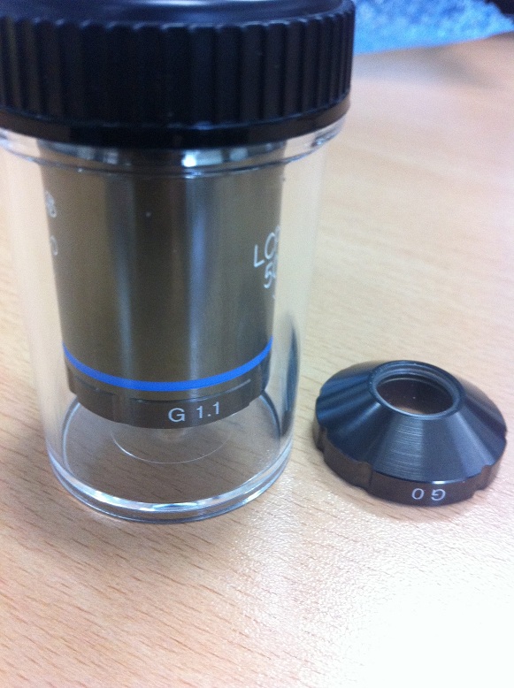
LCPLAPO50X
Profile of Jin Jae-Hwan ( Instructor )
CEO of JNOpTIC co., ltd
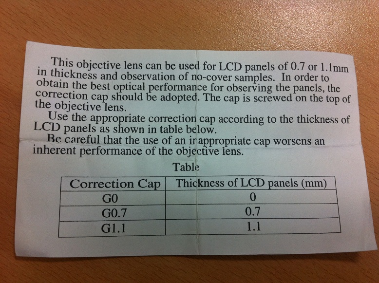
June 25, 2021
Patent registration:
Observation method and observation device
Simultaneous observation
( Bright Field and Fluorescence microscopy)
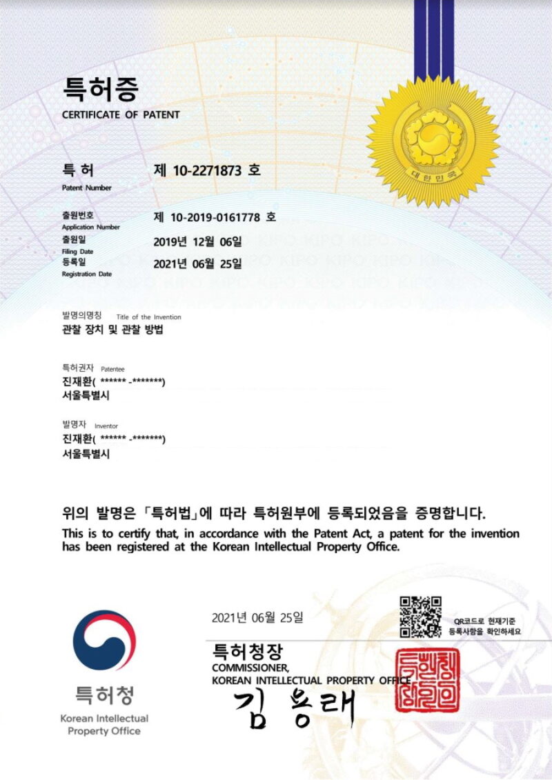
March 09, 2021
Patent registration:
sample fixing device and method of placing sample
Patent applied to section observation microscope
(JNO-FM-BX53-SET)
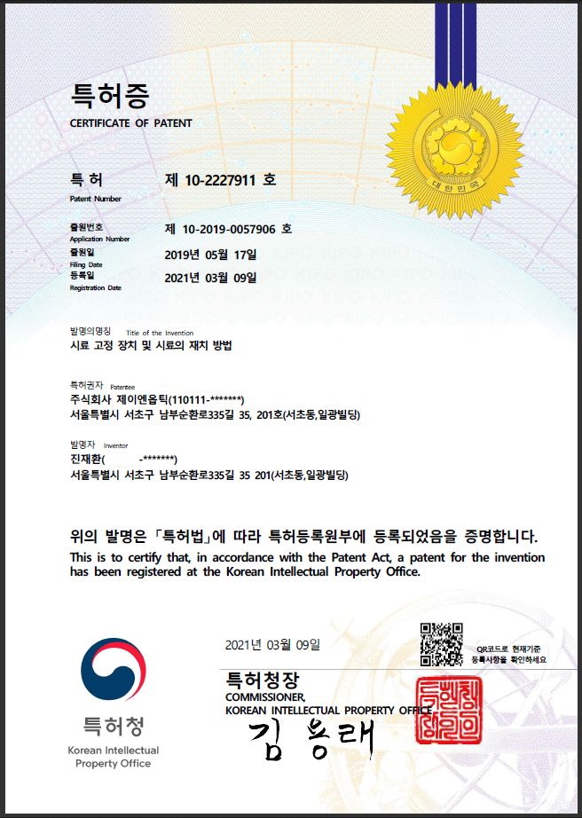
June 12, 2014
Patent registration:
Method of measuring the height of a sample using a microscope
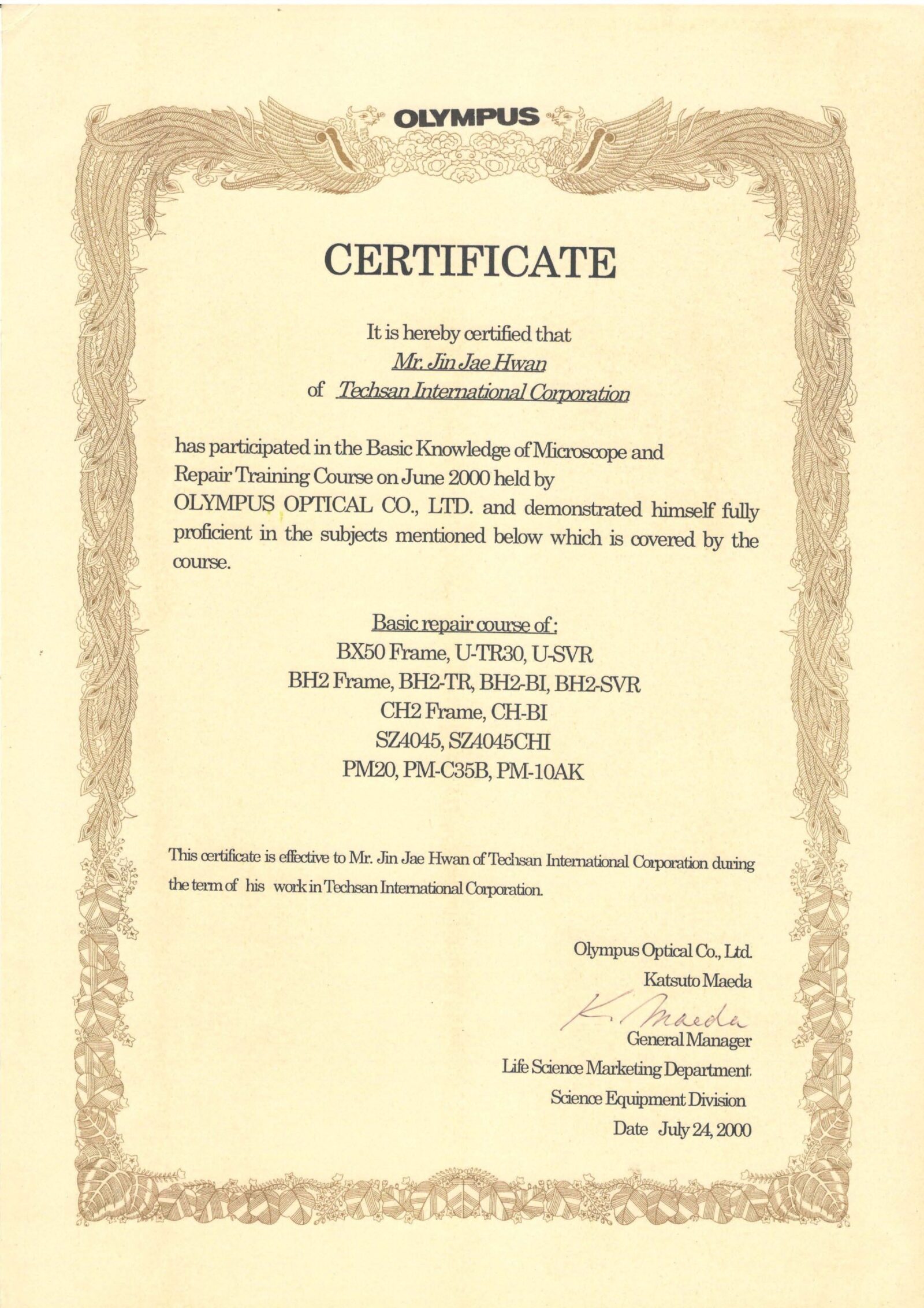
June 5th ~ June 15th, 2000 ( OLYMPUS Japan in Tokyo)
Basic Knowledge of Microscope and Repair Training Course
September 4, 2003 ( OLYMPUS Japan in Tokyo)
Measuring Microscope Calibration License
Main Model: STM6
License ID Number: M029
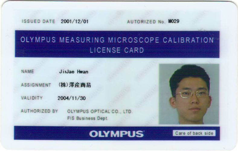

September 4, 2003 ( OLYMPUS Japan in Tokyo)
Biological Confocal Microscope Setup & Service Training
Main Model: FluoView300 & FluoView500
October 3, 2003 ( OLYMPUS Japan in Tokyo)
Inverted Microscope & Research Stereo Microscope Service Training
Main Model: IX71 / SZX9
August 17th ~ August 20th, 2004 ( OLYMPUS Japan in Tokyo)
Confocal Microscope Setup & Service Training
Main Model: FV1000
October 11 ~ October 13, 2004 ( OLYMPUS Japan in Tokyo)
Confocal Microscope Setup & Service Training
Main Model: FV1000 M-COMB(Multi Combiner)
June 20, 2005 ~ June 23, 2005 ( OLYMPUS Japan in Tokyo)
Research System Microscope Service Training
Main Model: AX70
August 1st-August 2nd, 2005 ( OLYMPUS Japan in Tokyo)
Polarizing Microscope Instruction Training
Main Model: BX51P
December 19, 2005 ( OLYMPUS Japan in Tokyo)
ZDC Training for Maintenance & Basic Knowledge of Confocal Microscope
December 20 ~ 22, 2005 ( OLYMPUS Japan in Tokyo)
Brushup Course to bring out the power of a microscope (GA Academy)
August 2nd ~ August 4th, 2006 ( OLYMPUS Japan in Tokyo)
Confocal Laser Scanning Microscope for Industrial Market
D class License(for ols3000)
Main Model: OLS 3000(Lext)
October 11 ~ October 15, 2006 ( OLYMPUS Japan in Tokyo)
Confocal Laser Scanning Microscope Training for Bio Maket
Maintenance Service Training
Main Mode : M-COMB(Multi Combiner for FV1000)
December 9, 2006
IX Repair training
May 07 ~ May 11, 2007 ( OLYMPUS Japan in Tokyo)
Modify Training for Zero-Drift Compensation Unit of IX81
IX2-Cusominsing Training (Optic Port Modify)
Main Model: IX71 & IX81-ZDC
September 3rd ~ September 08th, 2007 ( OLYMPUS Japan in Tokyo)
Leicense D class Trainging
Main Model: OLYMPUS Bio Confocal Microscope FV1000
April 08 ~ April 10, 2008 ( OLYMPUS Japan in INA_Nagano )
IV100 Setup Training
(in vivo fluorescence molecular imaging systems)
July 22 ~ July 23, 2008 ( Narishige Group in Tokyo, Japan )
Narishige Maintenance Training
July 24th ~ July 28th, 2008 ( OLYMPUS Japan in Tokyo)
Research Inverted Microscope maintenance Training
Main Model: IX81
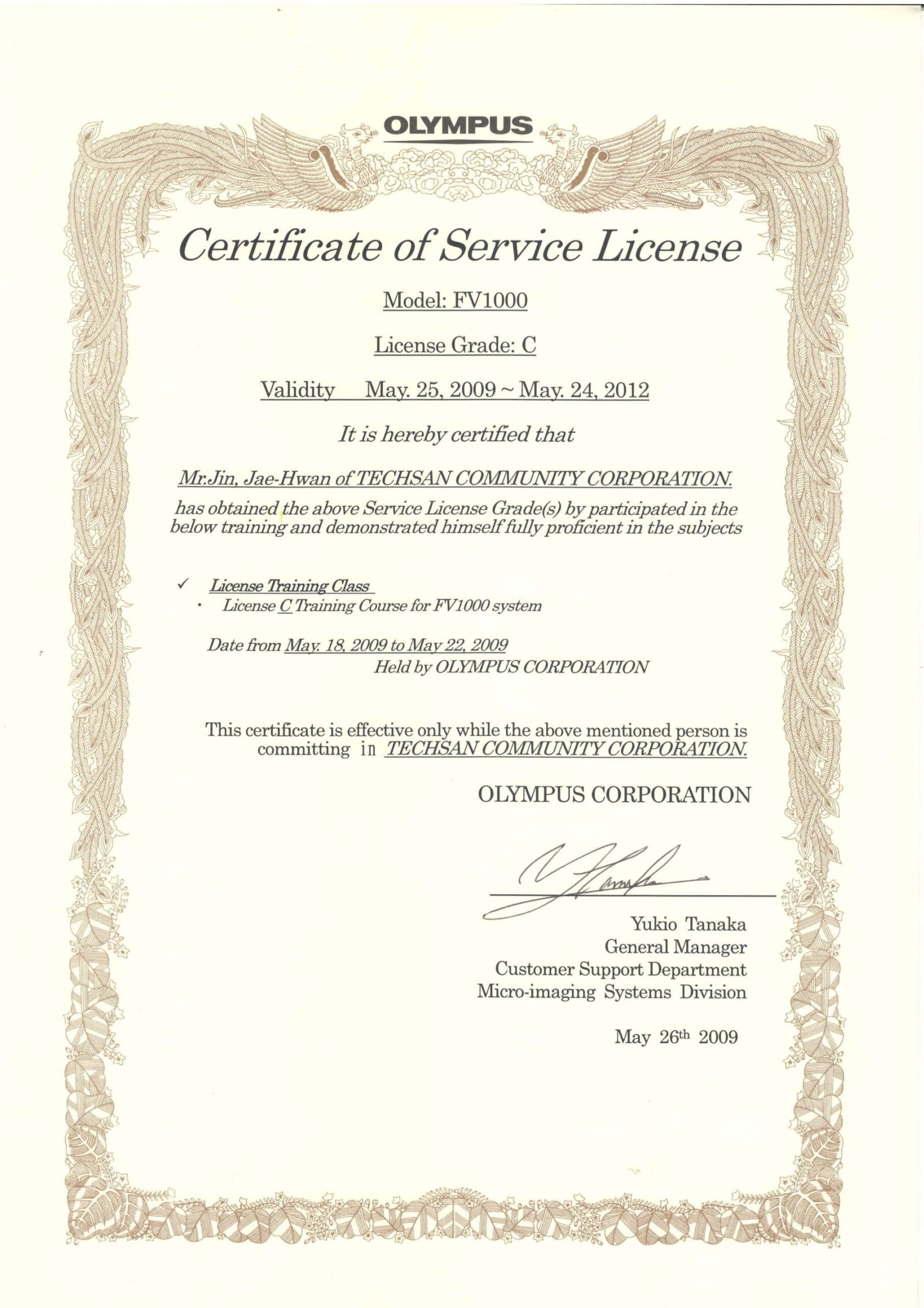
May 18 ~ May 22, 2009 ( OLYMPUS Japan in Tokyo)
OLYMPUS Bio Confocal Microscope License Training
License Grade: FV1000 C class
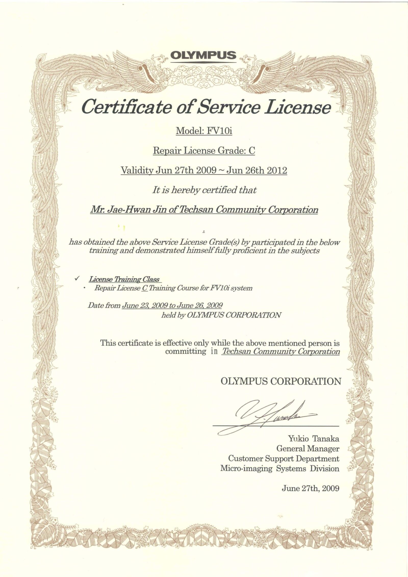
June 23 ~ June 26, 2009 ( OLYMPUS Japan in Tokyo)
Confocal Laser Scanning Microscope License Training
License Grade: FV10i C class
October 19 ~ October 23, 2009 ( OLYMPUS Japan in Tokyo )
Multi Photon Laser Scanning MICROSCOPE Setup Training
Main Model: MPE C class
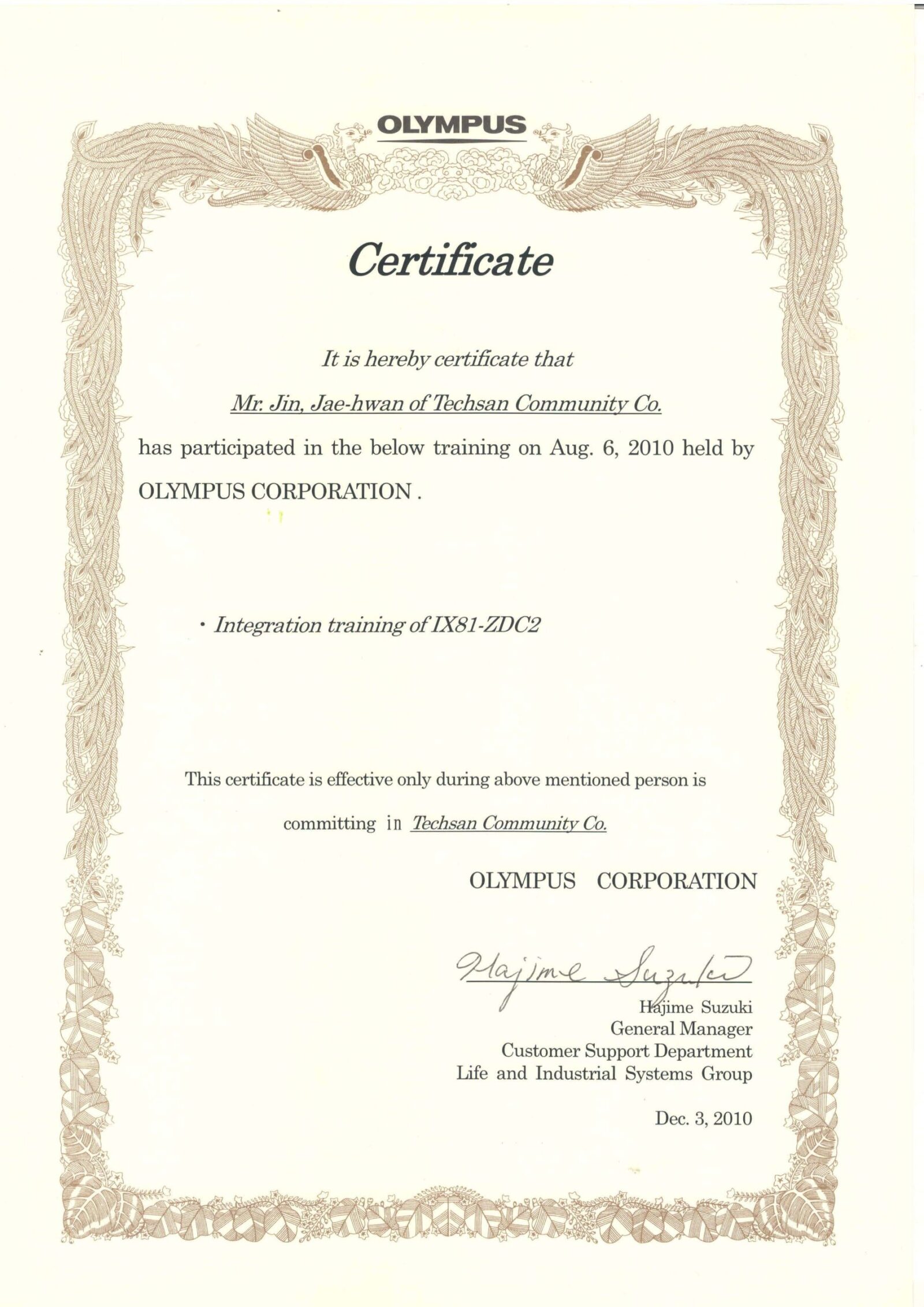
August 3rd ~ August 8th, 2010 ( OLYMPUS Japan in Tokyo )
Modify Training for Zero-Drift Compensation Unit of IX81
Main Model: IX81-ZDC2
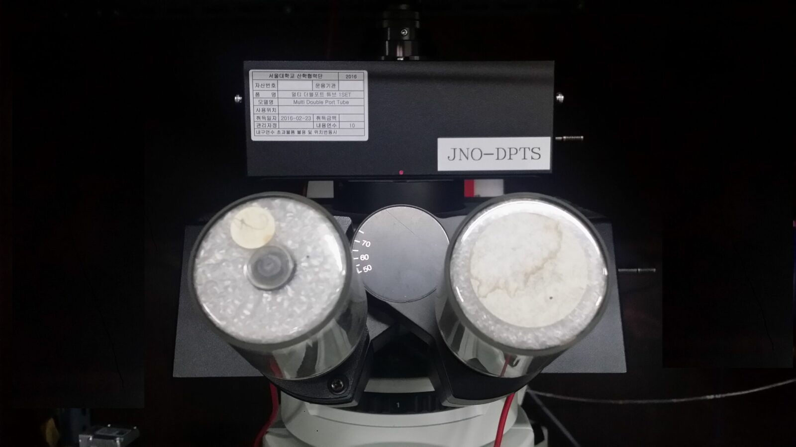
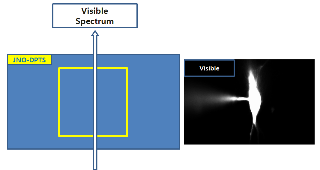
JNO-DPTS (Down load Catalog JNO-DPTS)
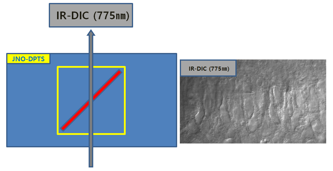
The Unit to observe fluorescence image and IR DIC image at the same time with Multi-Dual Port tube of BX51-WI
(Made by JNOpTIC co.,ltd )
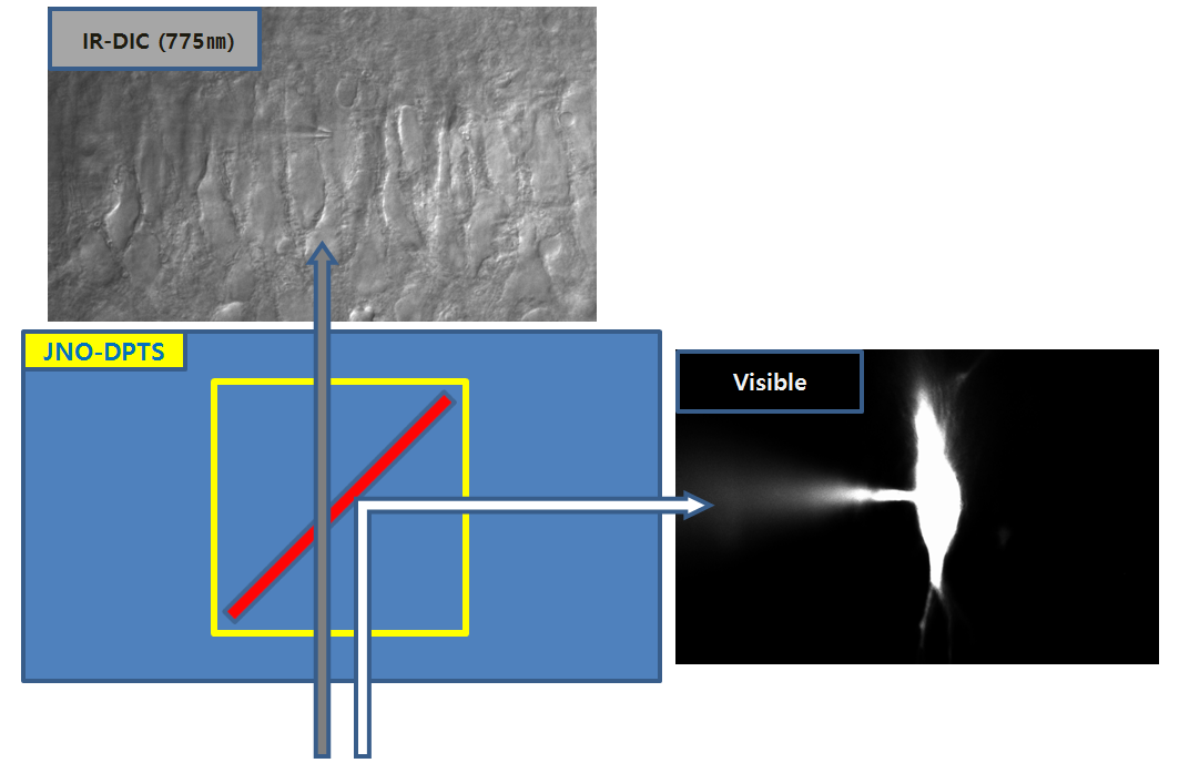
2-1. One Way((IR-DIC 775nm)
2-2. One Way((Visible Spectrum)
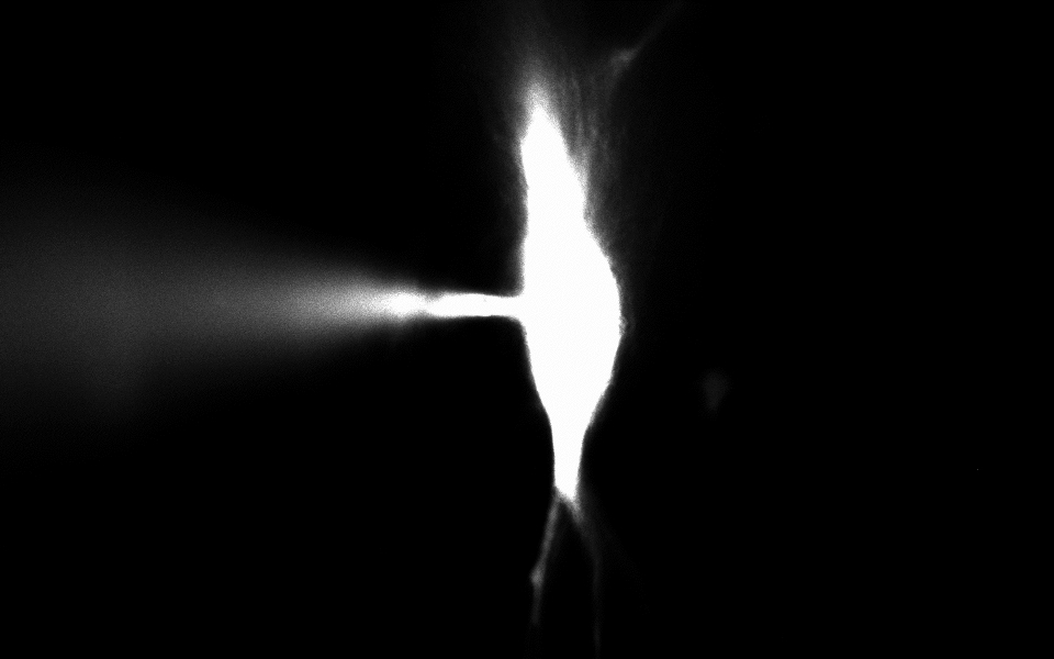
Simultaneously observation
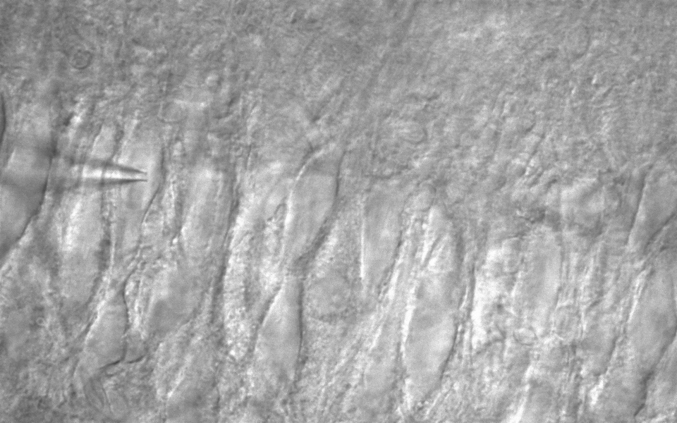
– CA3 pyramidal neuron, IR-DIC & Alexa Fluor 488 (50 uM)
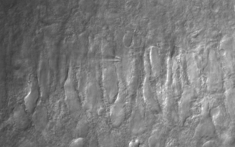
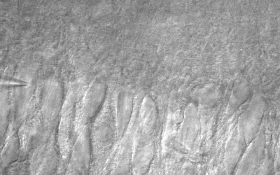
Advance Realtime Monitor. Easy-to-Use, Image Enhancement, etc
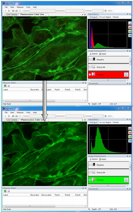
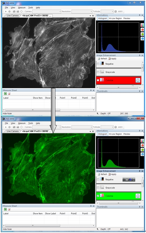
ARM is image analysis software for JNOPTIC AcquCAM cameras. This S/W is interchangeable with all WDM cameras regardless of camera brand and model and even more the most strength point is simple and easy use of length, area, angle, etc.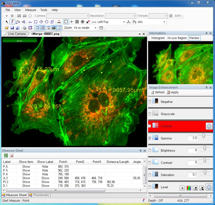


Improvement Function about Microscopy Image Monochrome. (Used function: Auto Level)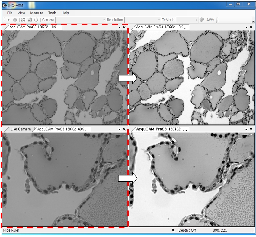 Effective improvement of image with simple operation. This image is shot by JNOPTIC Pro/S3 camera
Improvement Function about Microscopy Image. (Used function: Auto Level)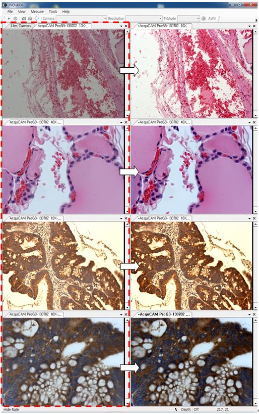 Effective improvement of image with simple operation. These images are shot by JNOPTIC AcquCAM Pro/G3 camera
| Responding Model | Measurement value unit | Measurable height | Error of measuring 1000㎛ | Recommended measuring height |
| CX 41 | 0.2 ㎛ | -9.9 ~ 29.9㎜ | Below ±20㎛ | Below ± 2000㎛ |
| CKX 41 | 0.2 ㎛ | -9.9 ~ 29.9㎜ | Below ±20㎛ | Below ± 2000㎛ |
| BX – FM | 0.2 ㎛ | -9.9 ~ 29.9㎜ | Below ±20㎛ | Below ± 2000㎛ |
| BX 51/53 | 0.1 ㎛ | -9.9 ~ 29.9㎜ | Below ±10㎛ | Below ± 1000㎛ |
| MX 51 | 0.1 ㎛ | -9.9 ~ 29.9㎜ | Below ±10㎛ | Below ± 1000㎛ |
| MX 61L/61 | 0.1 ㎛ | -9.9 ~ 29.9㎜ | Below ±10㎛ | Below ± 1000㎛ |

JNOPTIC Image Analysis Software OpTIC Eye Series
기본측정+오토카운팅(입도분석용)+금속조직분석용
– 형상측정(상분율측정)
: 두가지 이상의 색상을 추출하여 면적율 계산
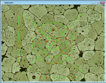
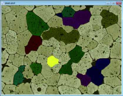
– 그레인사이즈(Grain Size Analysis)
: ASTM E 1382에 의한 금속표면의 결정립도 산출
: 교차점 / 면적 / 수동면적 추출방식
– 구상화율 분석(Nodular-graphite analysis)
: ASTM, ISO, JIS 규격에 따라 분석이 가능
– 비금속 개재물(Non-Metallic Inclusion)
: 산화물, 규산물, 황화물, 내화물, 광재등 추출
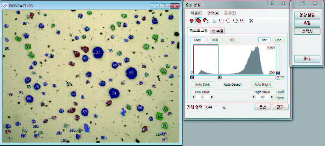
영상분석에 의한 구상화율 측정 (nodular-graphite analysis)
주철분석 소프트웨어 모듈로 ASTM, JIS, KS 규격에 따라 구상흑연주철의 구상흑연에 대하여 현미경영상분석을 통해 구상화율을 측정하고 결과를 표시합니다.
영상분석에 의한 결정도립 측정 (rain Size Analysis)