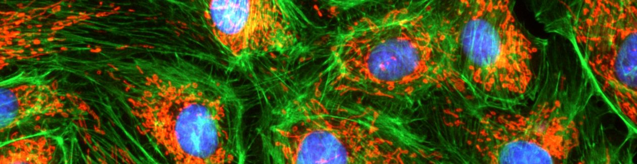
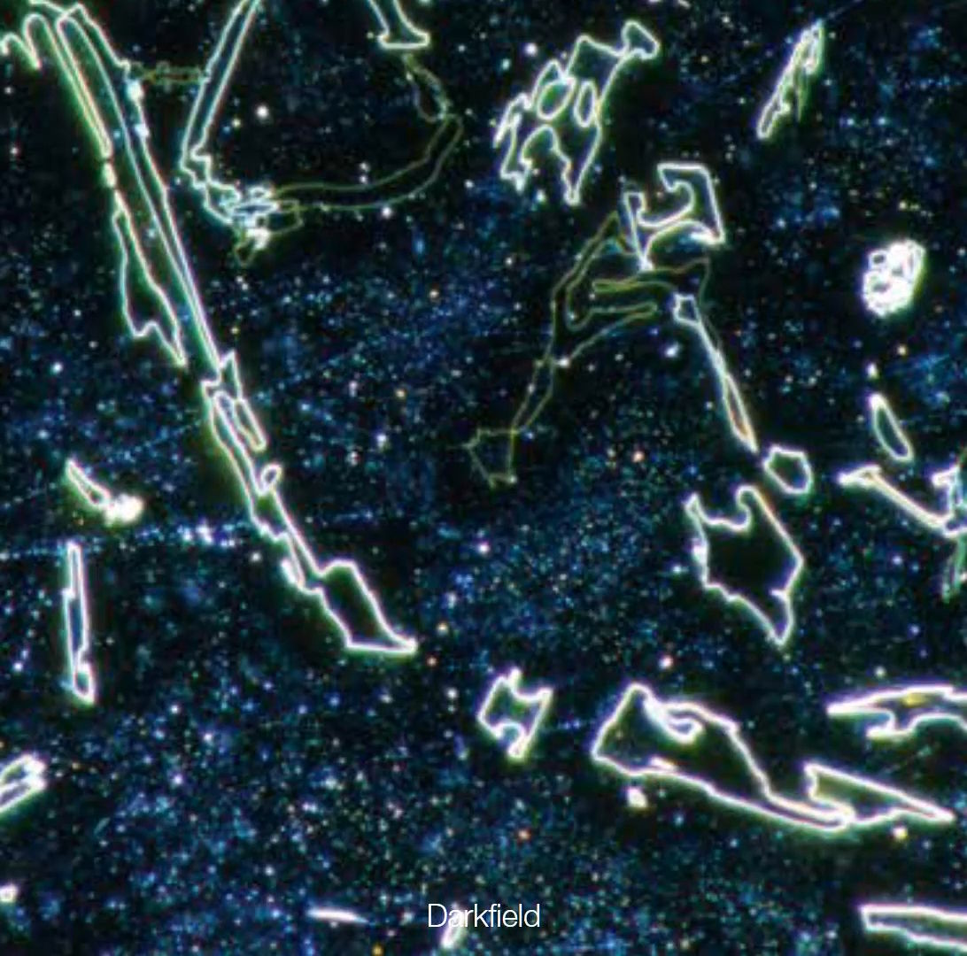
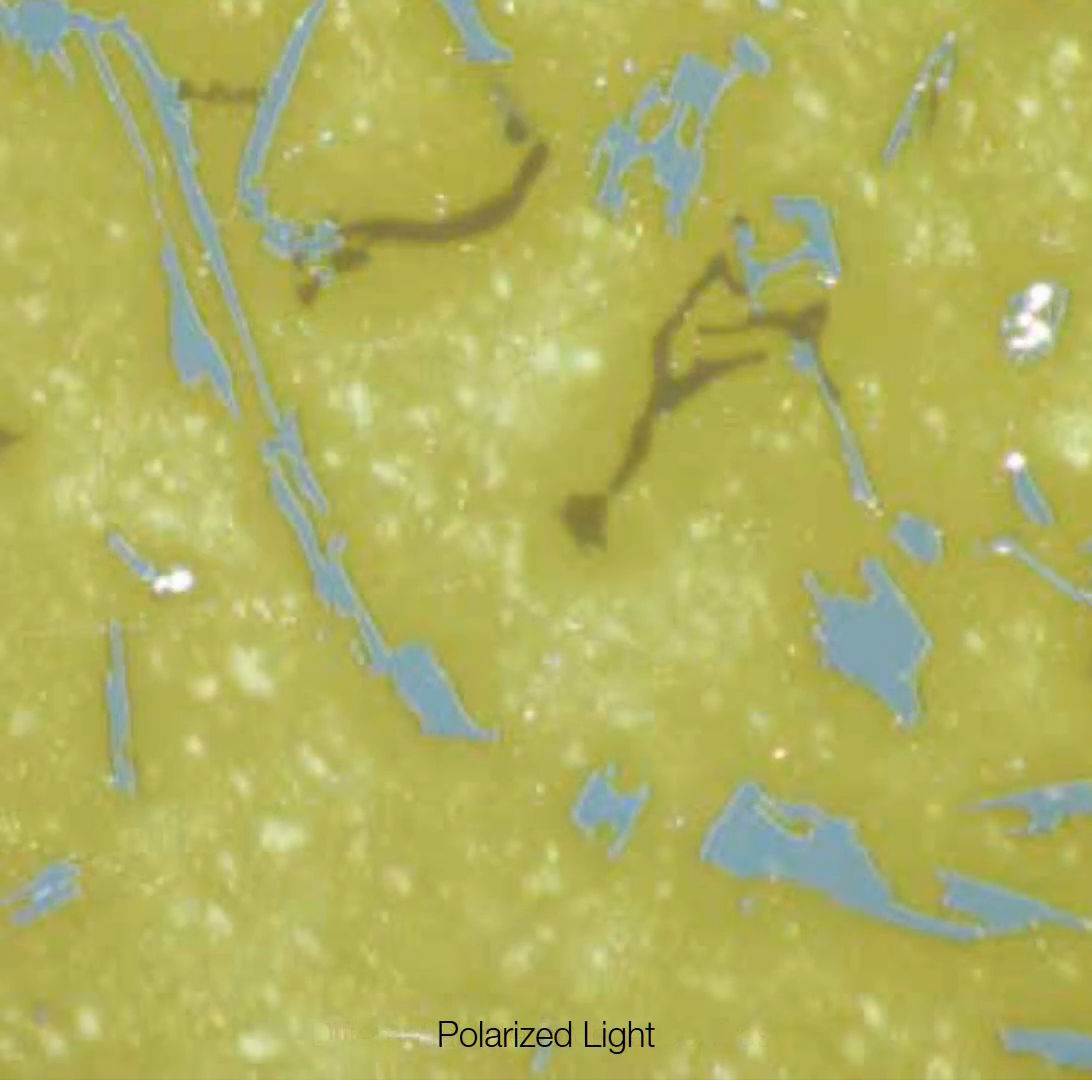
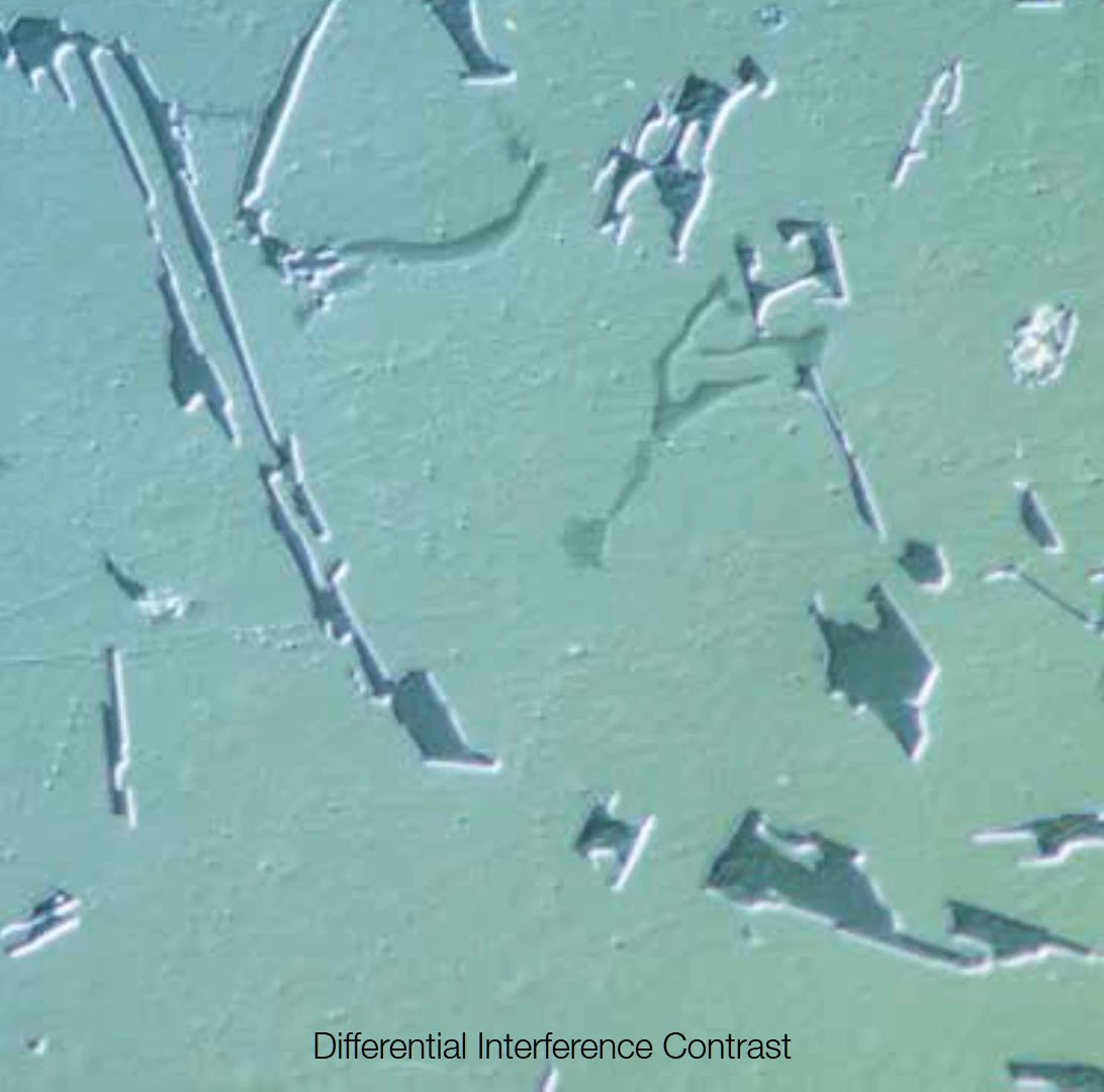
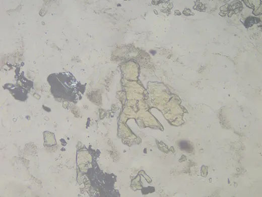
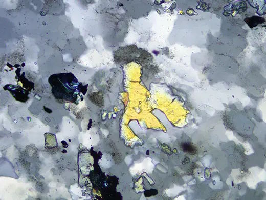
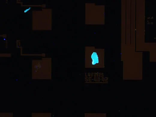
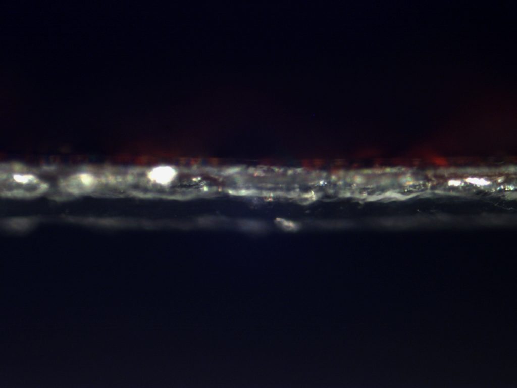
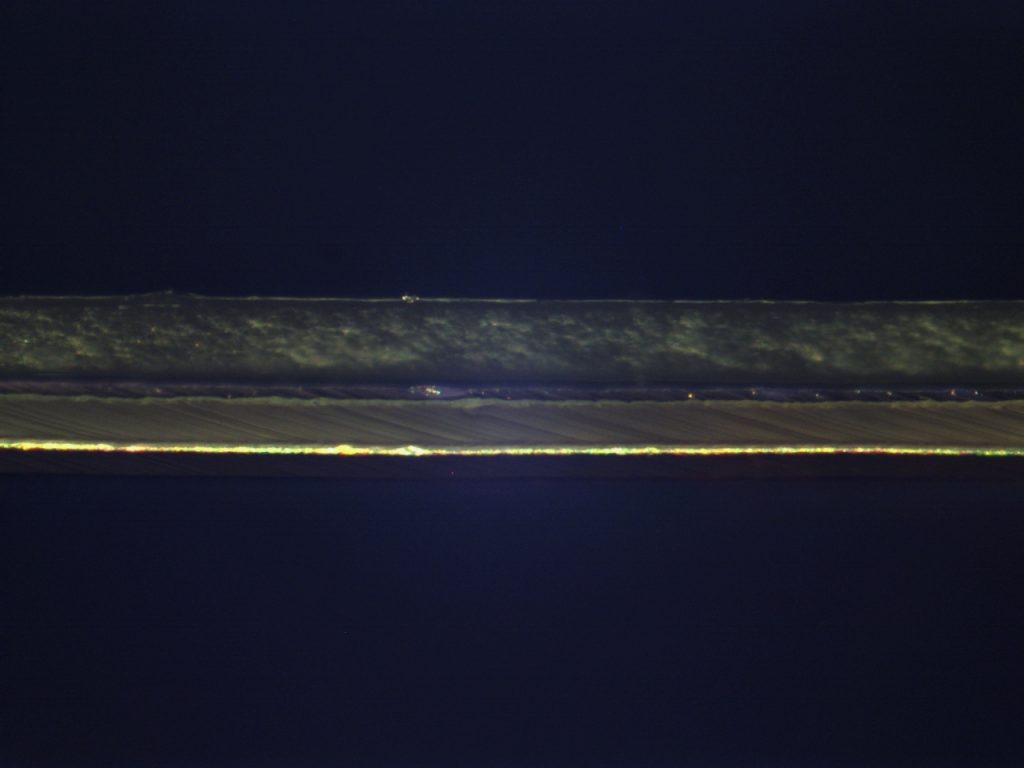
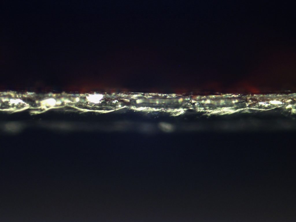
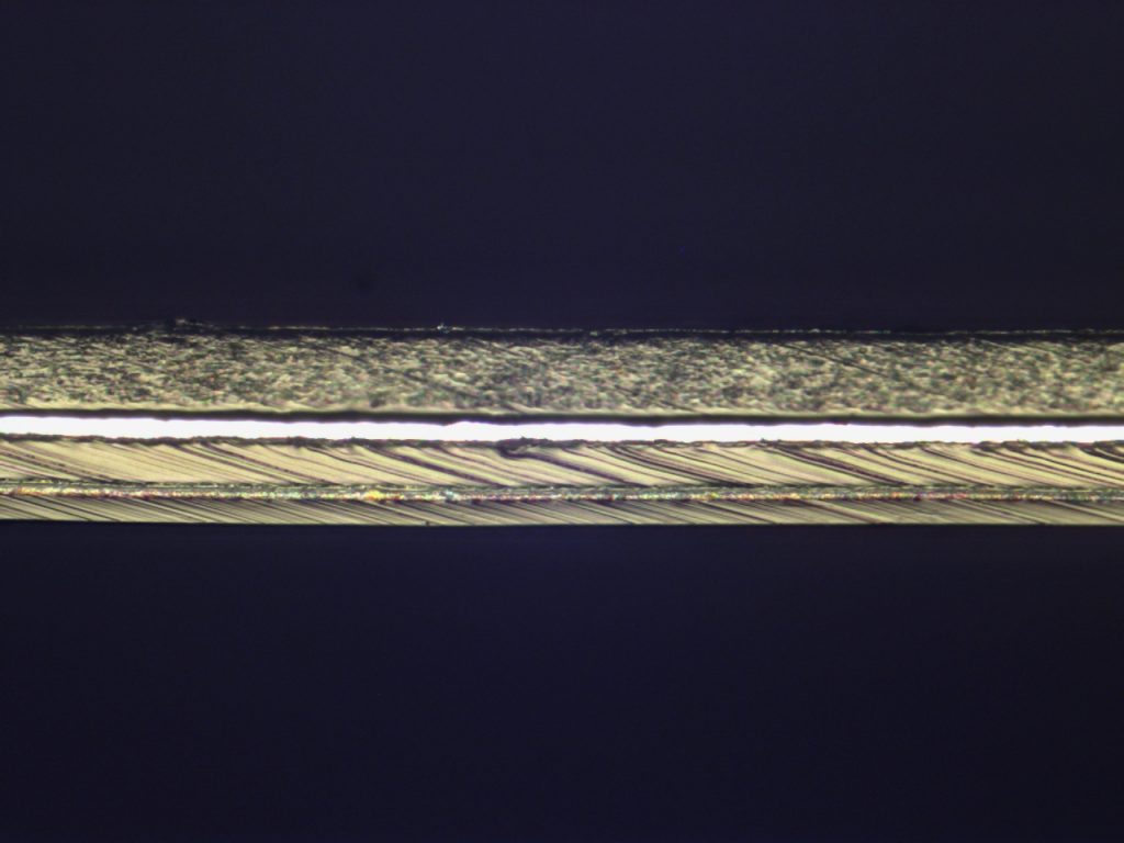
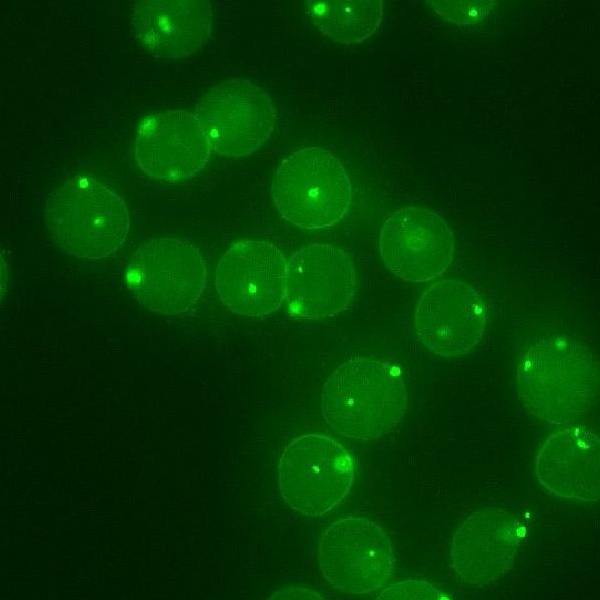
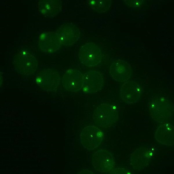
Bright Filed Image with BX53M Industrial_microscopeDark Filed Image with BX53M Industrial_microscopeSimple Polarized Image with BX53M Industrial_microscope (Simple Polarization)DIC Image with BX53M Industrial_microscope반사형 현미경 (Reflected Microsocpe) 검경법_명시야, 암시야, 간이편광, 미분간섭관찰(DIC)BF (구상흑연주철 )편광 (구상흑연주철 )Fluorescence image구상 흑연 주철의 명시야, 간이편광 이미지 와 형광이미지(우측) 입니다.
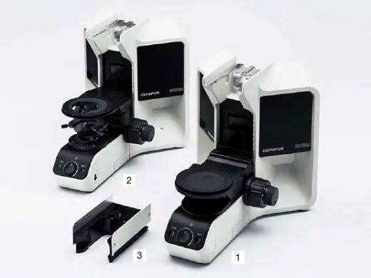
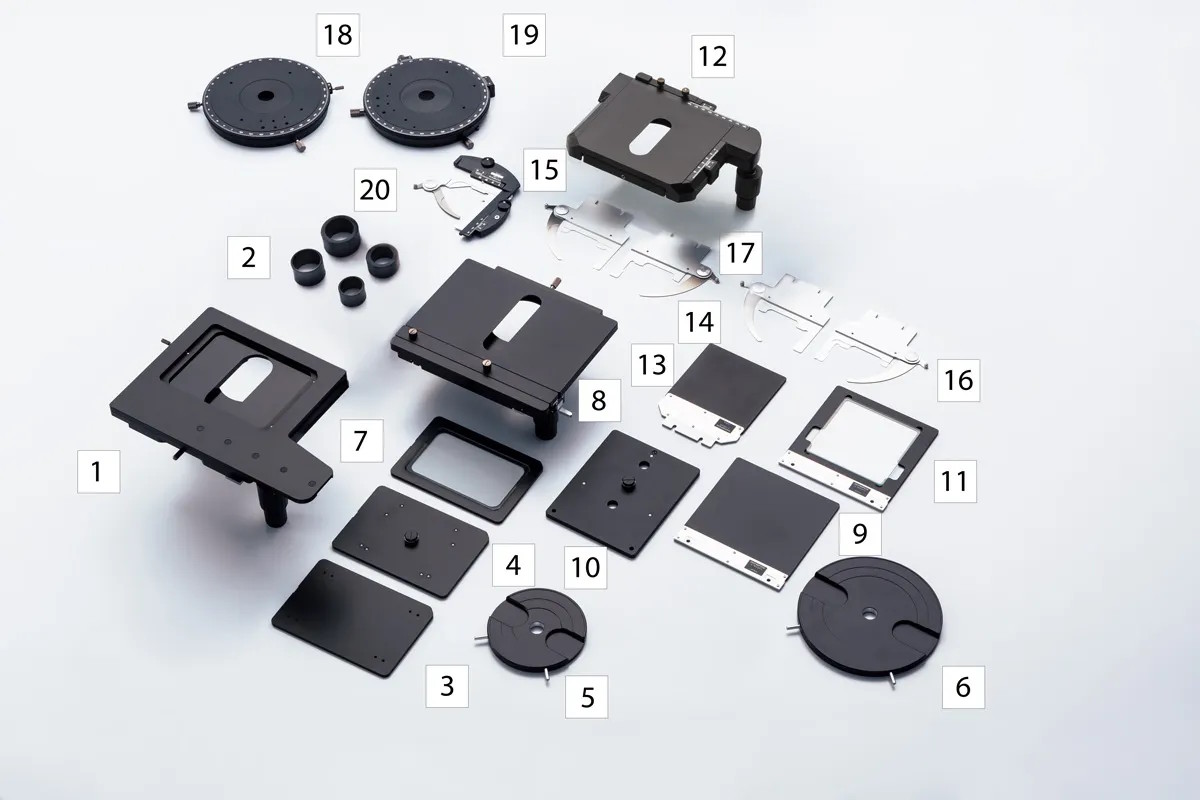
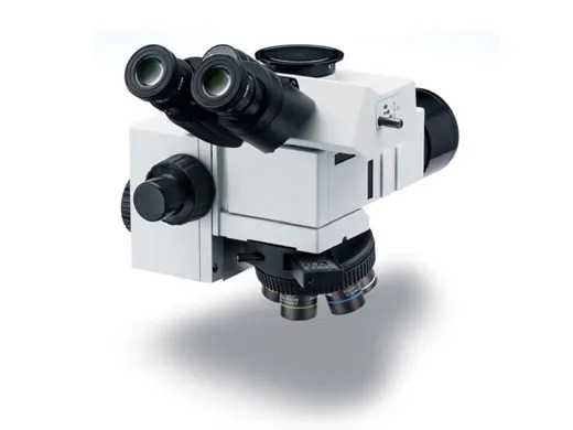
BX53M 구성을 위한 다양한 모듈( Module )
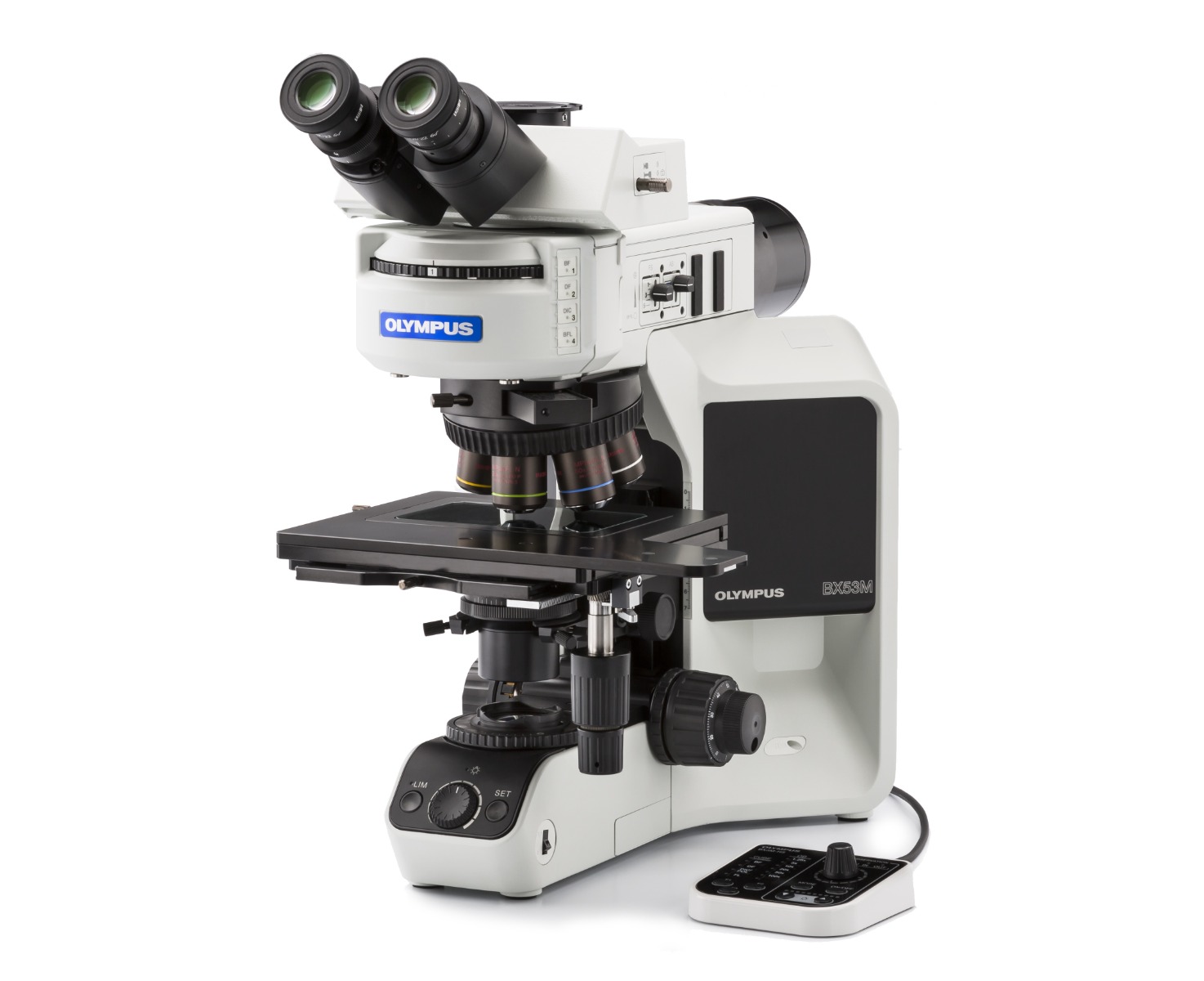
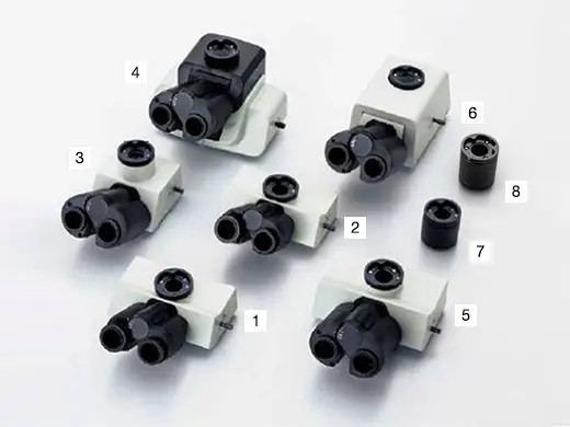
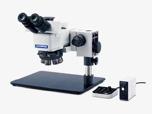
Body Frame for BX53MILLUMINATOR for BX53MHead for BX53MLight soruce for BX53MCondensor for BX53MC-mount adapter for BX53MStage for BX53M Industrial_microscope실험 목적에 적합한 모듈과 대물렌즈를 선택 하여 조립하시면 – BX53M 1Set – 가 됩니다 . 상기 이미지는 구매 희망자의 이해를 돕기 위한 것으로, 일부 모듈은 누락되어 있으니, 구매 하시기 전에 전문가의 상담을 받으시기 바랍니다.
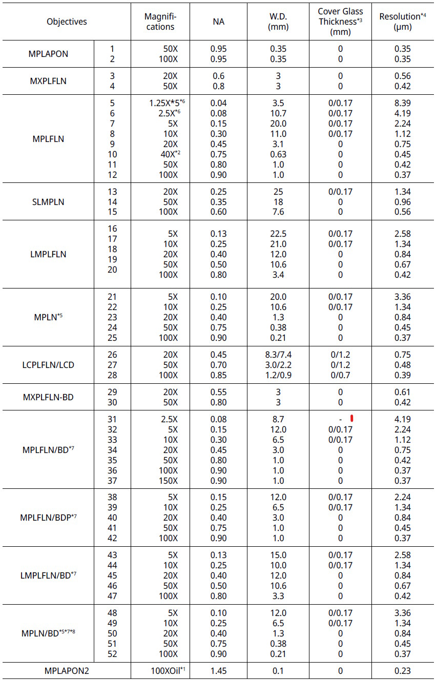
UIS2 Objectives ( 대물렌즈 )
UIS2 Objectives for BX53M Industrial_microscope
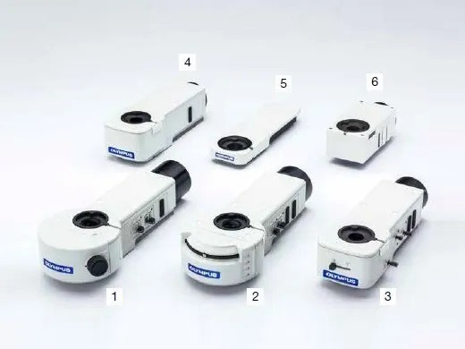
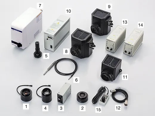
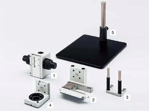
추가 유닛 ( Option )
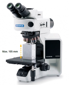
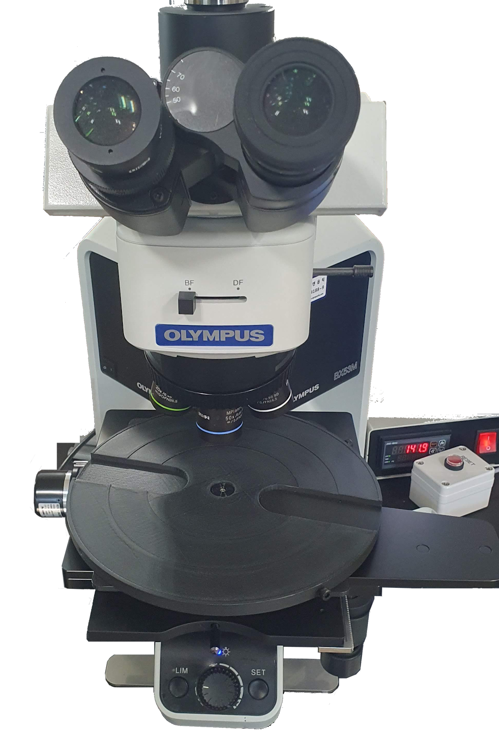
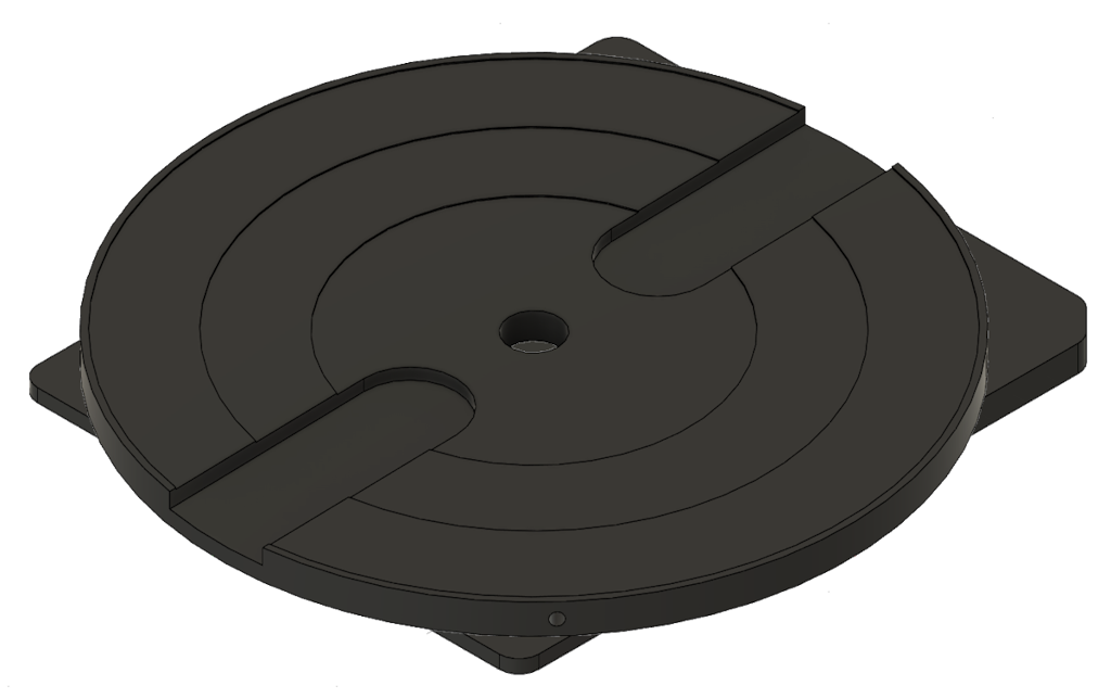
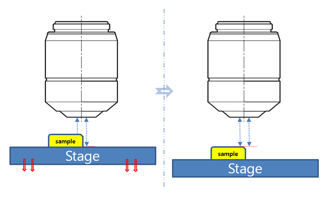
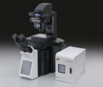
Flexibility for Sample Height and Weight
Body Frame 확장을 통한 최대 105mm(4.1인치)의 샘플을 스테이지에 올려서 검경이 가능합니다.
향상된 포커싱 메커니즘으로 최대 6kg 중량의 샘플을 올려 놓고 관찰 할 수 있습니다. (상기 중량은 스테이지의 중량이 포함된 값입니다. )
Flexibility for Sample Height and Weight Samples up to 105 mm can be mounted on the stage with the optional modular unit.
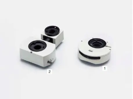
광로 변환 장치 및 추가 변배율 장치
Various types of accessories for multiple purposes. For use between tube and illuminator.
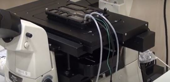
BXFM with BX3M-KMA-SBXFM with U-KMAS높이 측정 모듈 & Lagre StageBXFM은 한정된 BX53M의 고가의 Body Frame의 한계를 벗어난, 저렴한 Body 구성 뿐만 아니라, 다양한 관찰 환경에 맞추어 구성되어 판매 되고 있습니다.
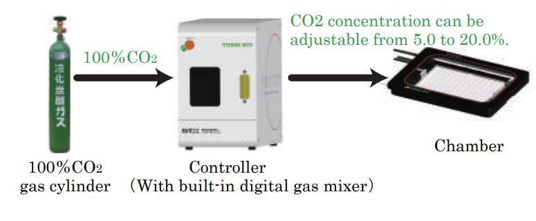
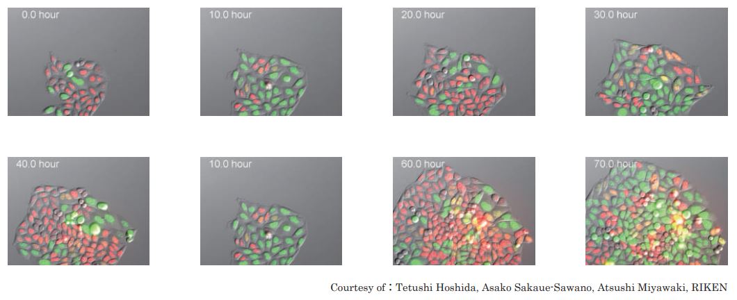
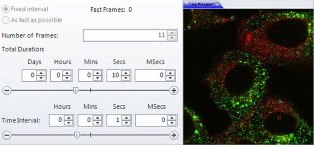
시간 간격으로 이미지를 획득하여 시간에 따른 표본 또는 재료의 변화를 조사합니다. 타임 랩스 이미지를 동영상으로 재생하여 움직임 및 기타 활동의 확인이 가능합니다.
시간변화에 따른 개체 이동변위 측정
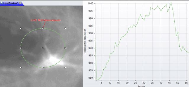
시간 변화에 따른 개체의 intensity 변화량 측정
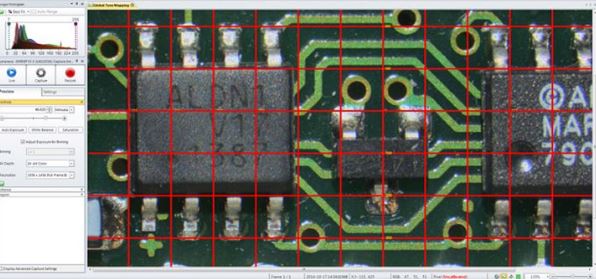
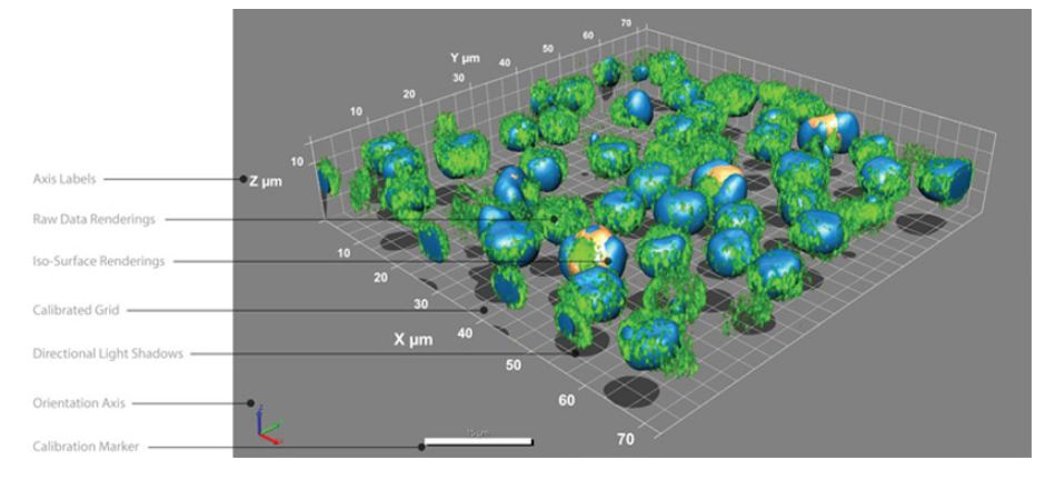
Morphology
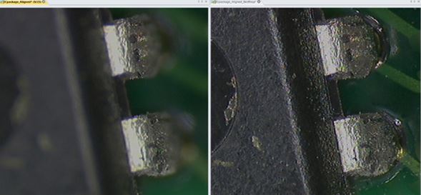
수동/ 자동 측정을 위한 다양한 필터를 사용하여 이미지 구조를 정확하게 분할합니다
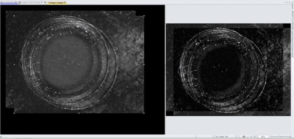
Filter and Enhance
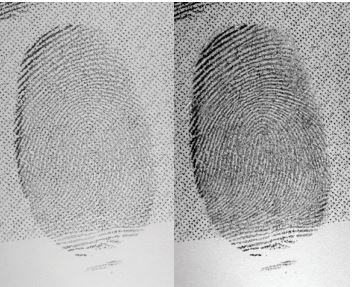
노이즈를 필터링하고 이미지 디테일을 향상시키기 위해 다양한 강화필터와 에지 필터를 사용합니다,
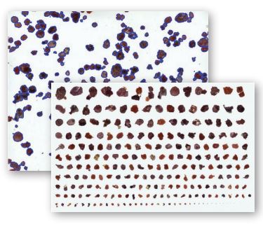
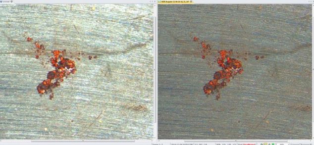
Pseudo-color
Pseudo-color(의사 색상)을 사용하여 회색조 이미지에서 관심있는 부분을 강조 표시합니다. 일반적으로 주변과 구분하기 어려운 특정 강도를 시각적으로 강조하기 위해 사용합니다.
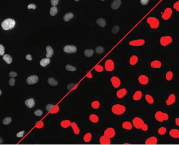
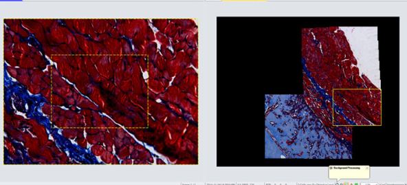
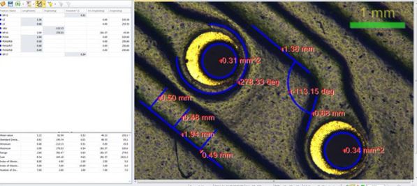
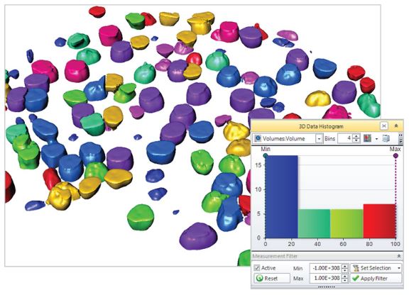
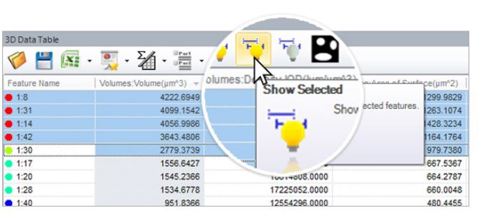
Count and Measure
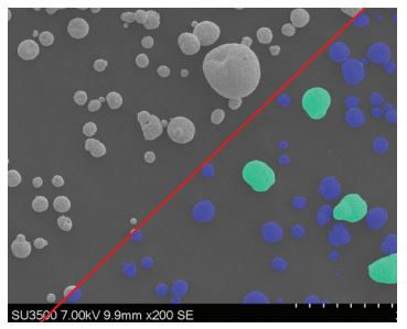
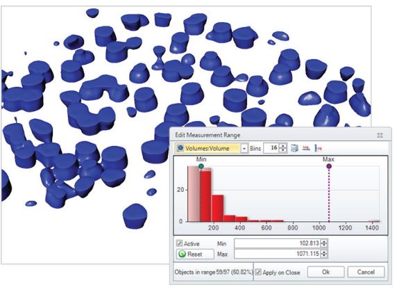
면적, 둘레, 길이, 진원도, 주축 및 부축, 각도, 중심, 내부홀 및 모집단 밀도등 50여가지가 넘는 수동 및 자동 측정 도구를 사용하여 각 객체를 분석합니다. 특정 객체에 태그를 지정하고 크기 또는 기타 측정 항목별로 정렬가능합니다
Count, Size & Sort Objects using Image-Pro Plus analysis software
Wound Healing Analysis – Image-Pro Plus Software
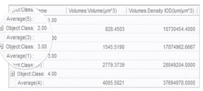
Classify
사용자 정의 분류 방법을 사용하여 분포 분석을위한 측정 매개 변수를 기반으로 셀, 입자 또는 객체를 클래스로 그룹화합니다
Threshold & Measure Objects
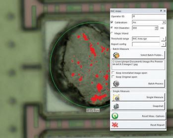
Interactive Measurements
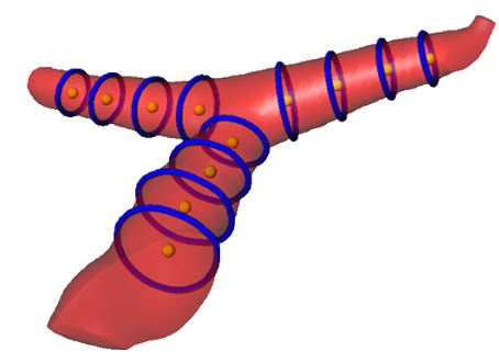
다양한 측정 옵션을 사용하여 이미지에서 수치화 가능한 데이터를 추출이 가능합니다. 계측 도구를 사용하면 가장 적합한 선, 호 및 원을 측정 할 수 있고. 캘리퍼 도구를 사용하여 를 규칙적인 간격 측정이 가능합니다.
Co-localization
생물학적 표본에서 공동 위치를 탐지하고 산점도에서 두 데이터 세트 간의 연관성을 그래픽으로 표시합니다.
Automate with Macros
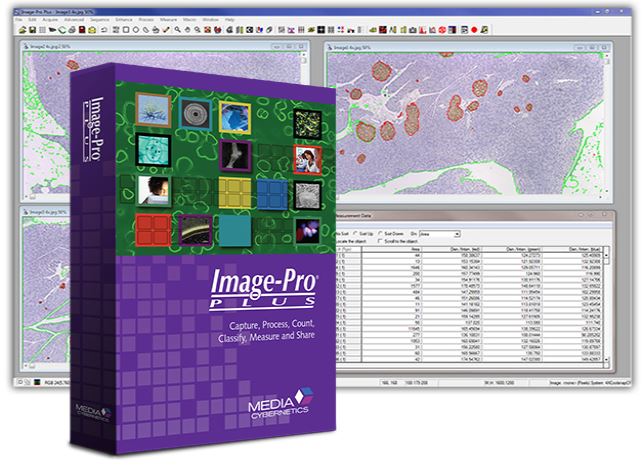
Image-Pro Plus는 편리한 사용자 정의 도구와 내장 매크로 프로그래밍 언어를 제공하여 이미지 분석 절차를 간소화합니다. macro 기록 도구를 사용하여 자주 수행하는 작업을 저장하고 쉽게 편집 할 수 있습니다. Image-Pro Plus에 포함 된 시간 절약형 macro를 사용하거나 사용자가 제공 한 Solutions Zone 사이트에서 다운로드 바랍니다.
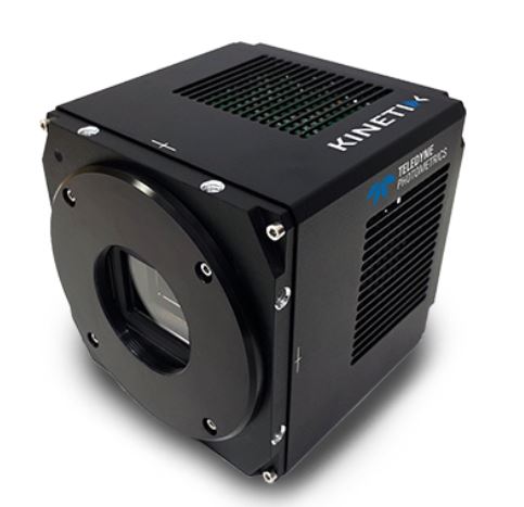
The brand new Kinetix family of back-illuminated sCMOS cameras delivers a framerate and field of view unmatched by any other sCMOS camera. KINETIX CAMERA