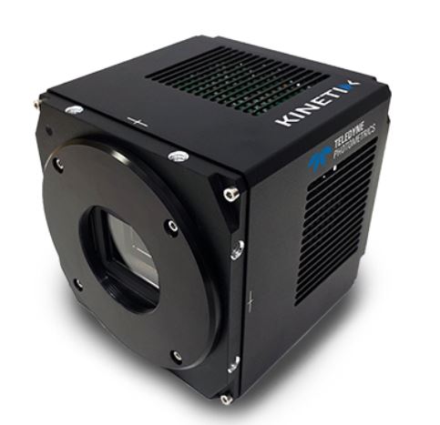
The brand new Kinetix family of back-illuminated sCMOS cameras delivers a framerate and field of view unmatched by any other sCMOS camera. KINETIX CAMERA
Patent: Matching the focus of the observation plane 필름단면의 초점 정합을 위한 장치 및 방법Patent: Height measurement method of sample by microscope
현미경을 이용한 샘플의 높이측정방법
Quad band catalog set for Multi LED light engines like those manufactured by Lumencor, CoolLED, Excelitas and Cairn Research. Excitation passbands are matched to discrete, triggerable LEDs appropriate for excitation of CFP/YFP/RFP/Cy7 and similar fluors. Not intended for use with white light sources.
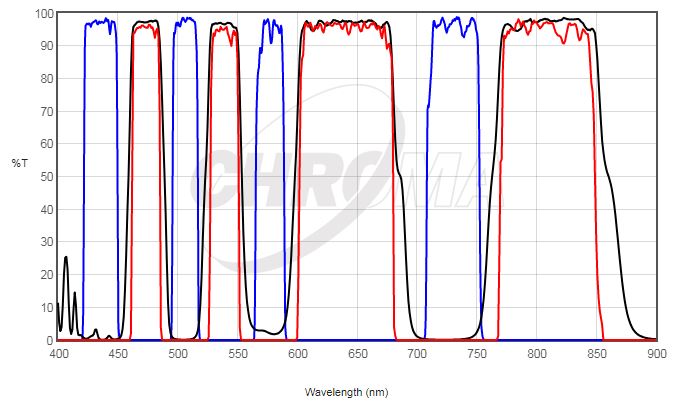
89403 Spectrum
chroma_89403
Filters
Type
89403x
EX
89403bs
BS
89403m
EM
89403 Spec
Typical Application(s) : Widefield Microscopy
Coating : Sputter/Hard Coated
Round excitation and emission filters mounted in anodized aluminum rings 2.3mm thick, up to 25mm in diameter (for standard sizes)
Rectangular dichroics up to 26x38mm, 1mm thick, to fit standard microscope manufacturer filter cubes (for standard sizes)
*** For filters larger than 25mm and dichroics larger than 26x38mm, please contact us for pricing.
NOTE: These require the use of filter wheels or sliders, or image-splitting devices to hold multiple individual filters.
Available of taking images with a wavelength of 650nm or more, such as CY5
High Sensitivity Sensor (Best)
Fast, seamless image transfer
Available of images with a very wide field of view
Low Noise Sensor
The displays live digital images with gradual smoothness and combines exceptional resolution with faithful color reproduction.The new sensors feature a global shutter function and able to capture a high-speed moving image without focal plane distortion. High-speed processing, low noise and low power dissipation by using column-parallel A/D conversion. equipped with trigger mode, and the external pulse can control accumulation time. The Sensor also have a pulse output function to indicate respective conditions during shutter operation and can be coordinated with peripheral circuits. High-definition images can be displayed live at a rate of high frames per second. without compression. Such imaging quality enables even the finest cellular regions to be observed clearly and distinctly without deterioration. While focusing is made stress free.
Images with AcquCAM 23GR
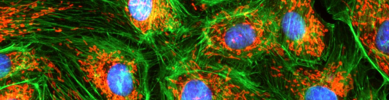
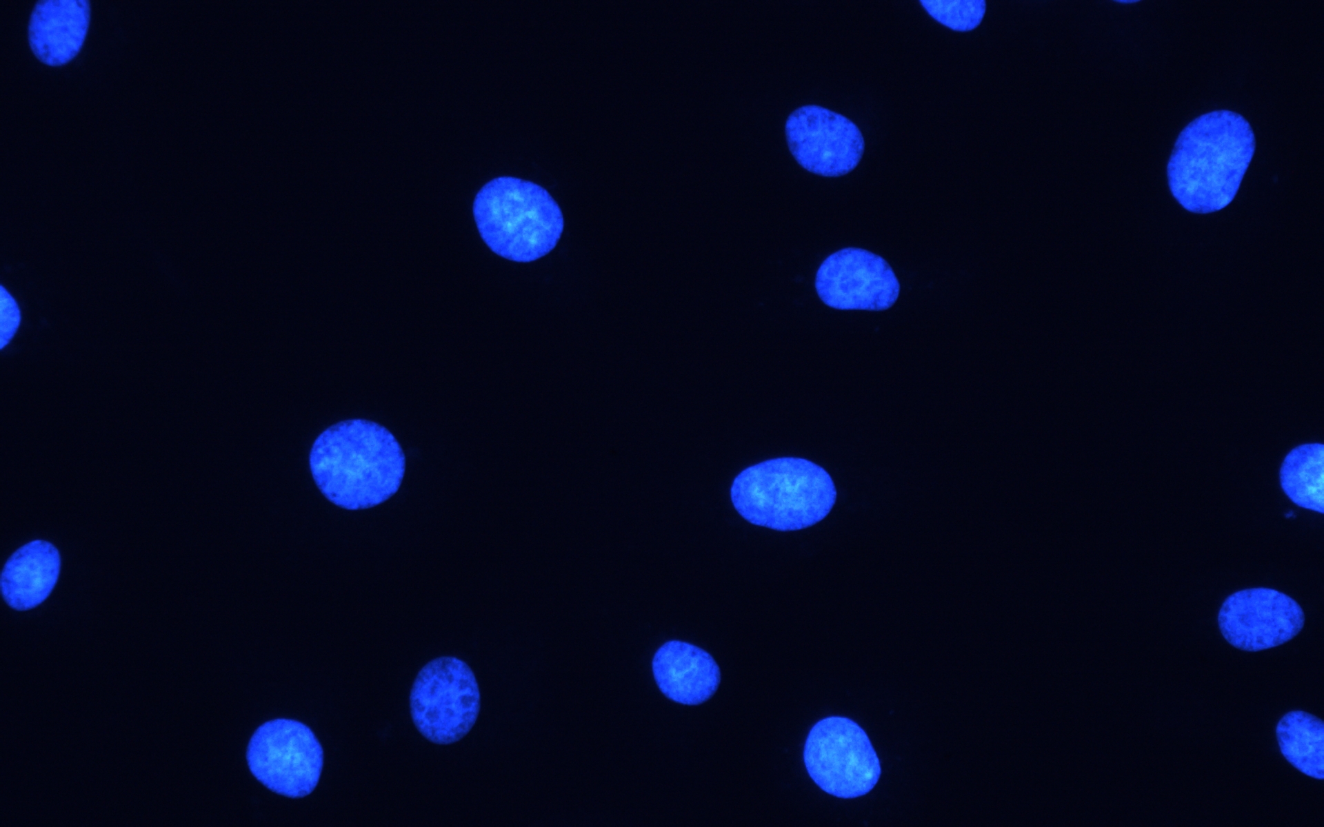
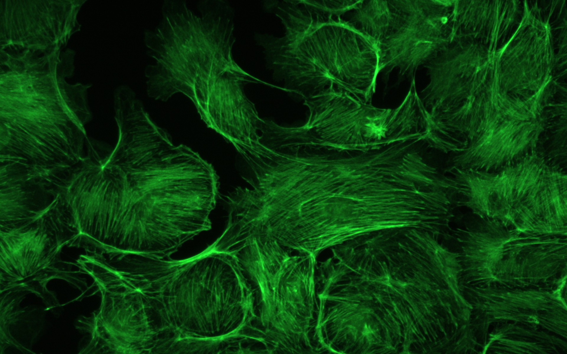
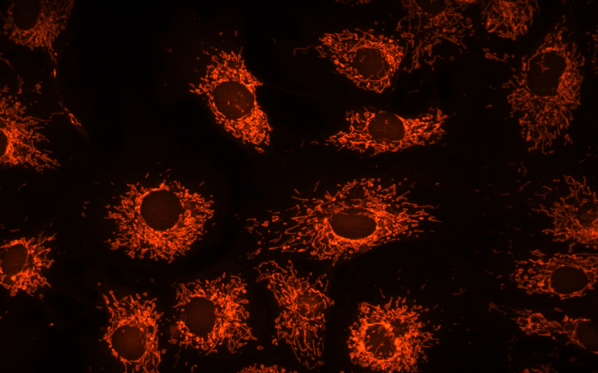
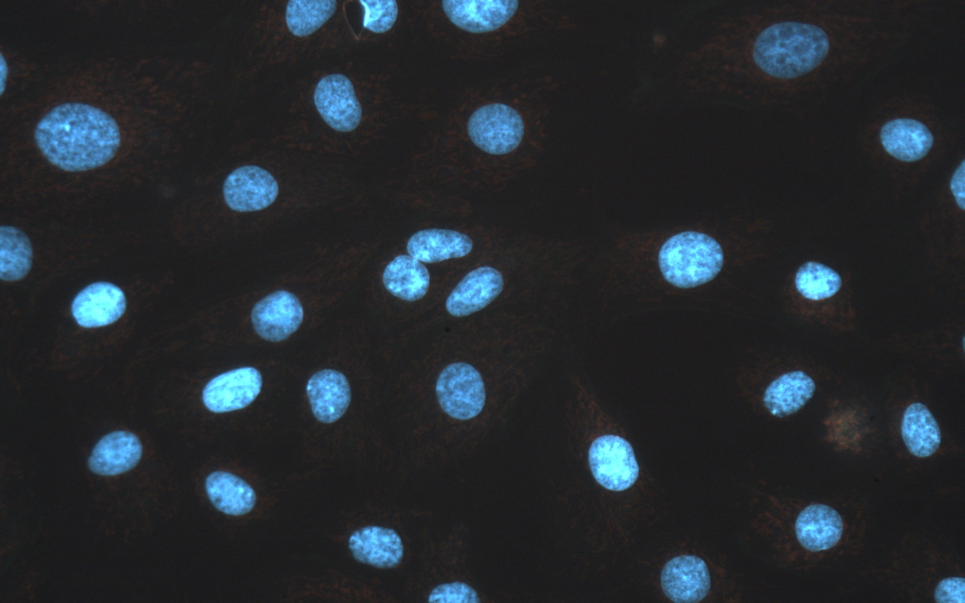
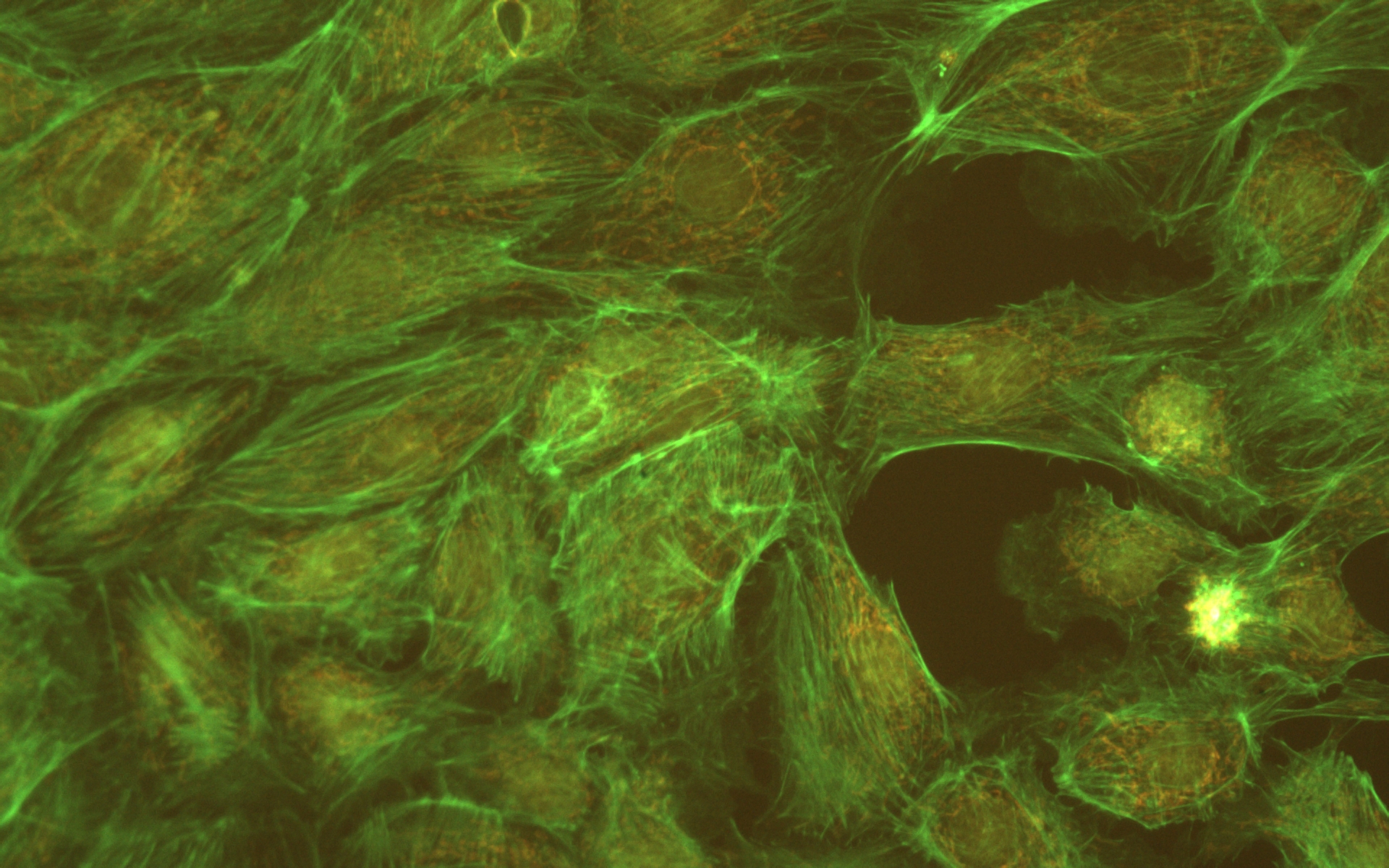
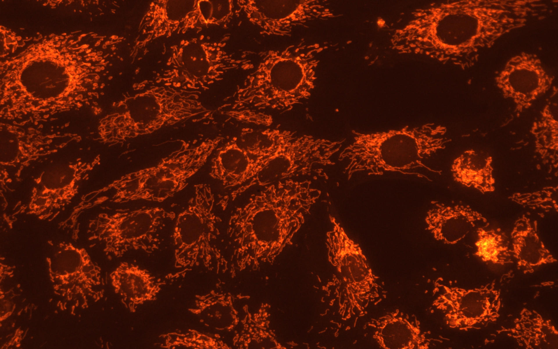
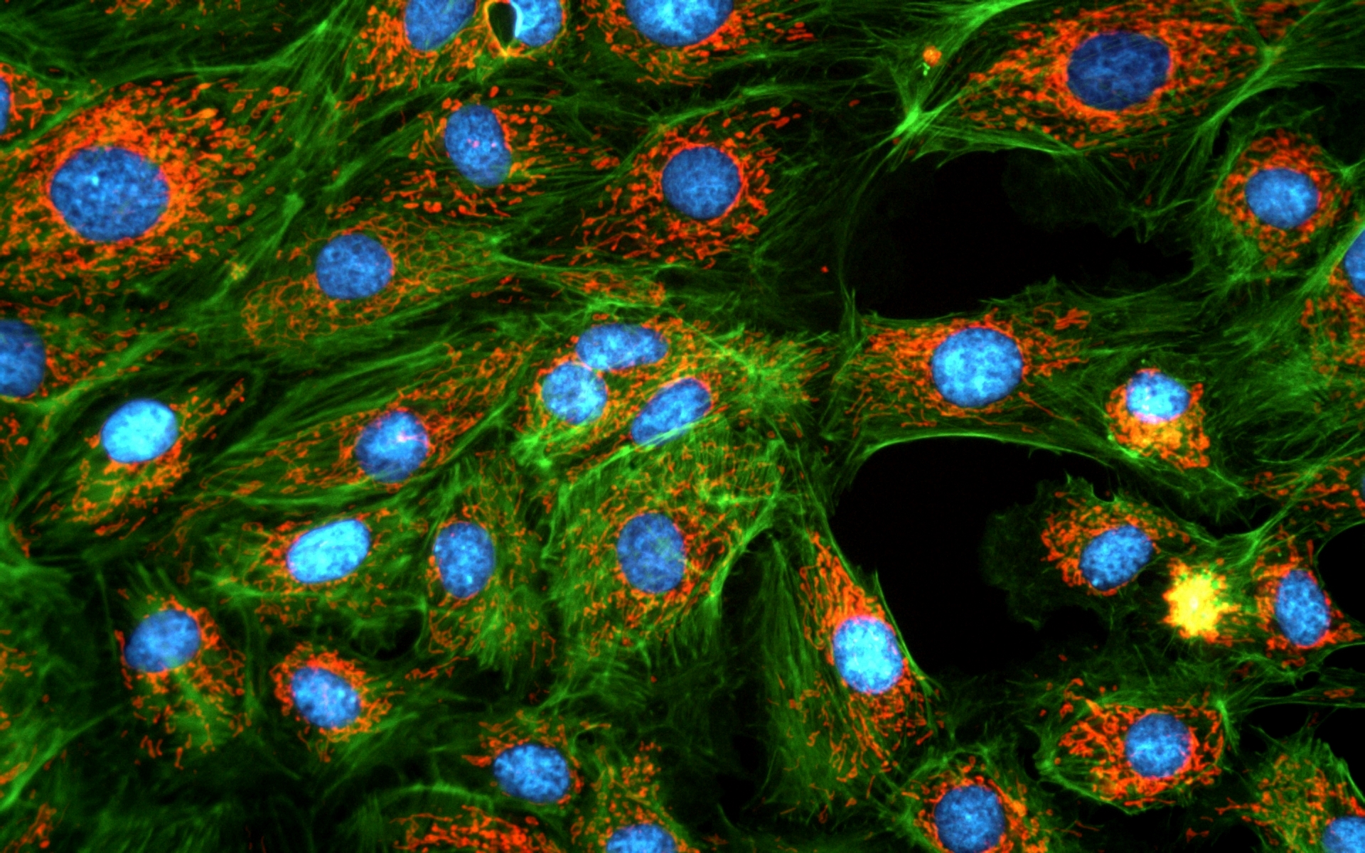
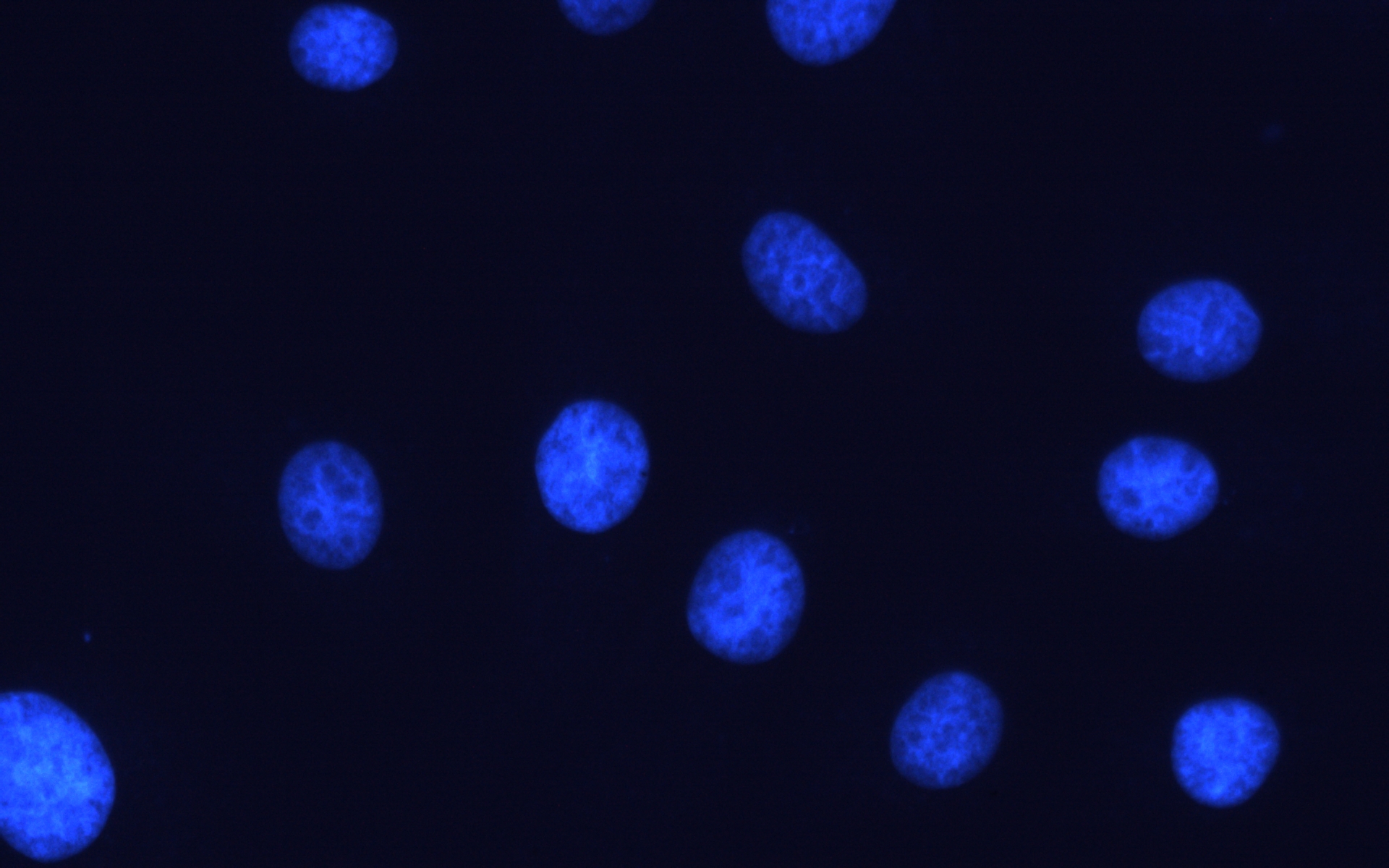
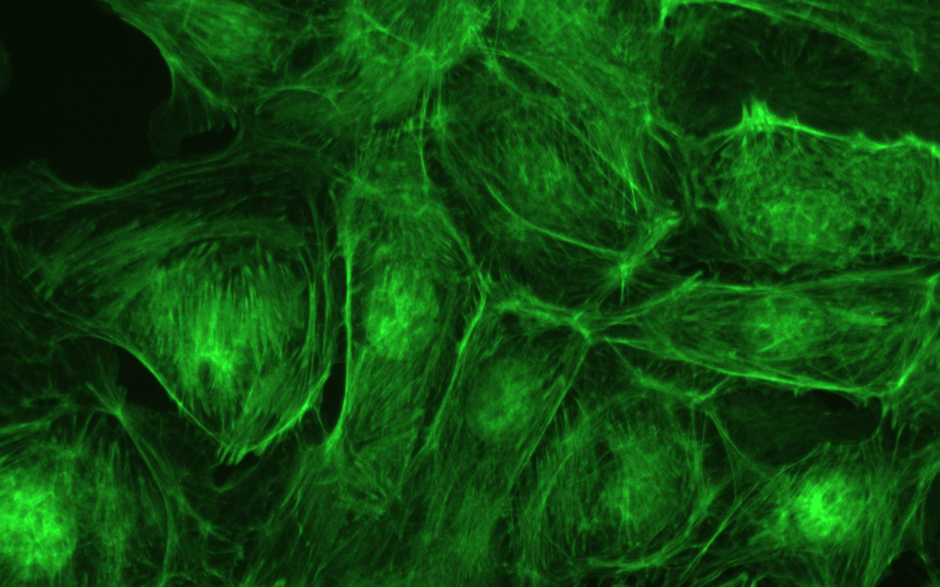
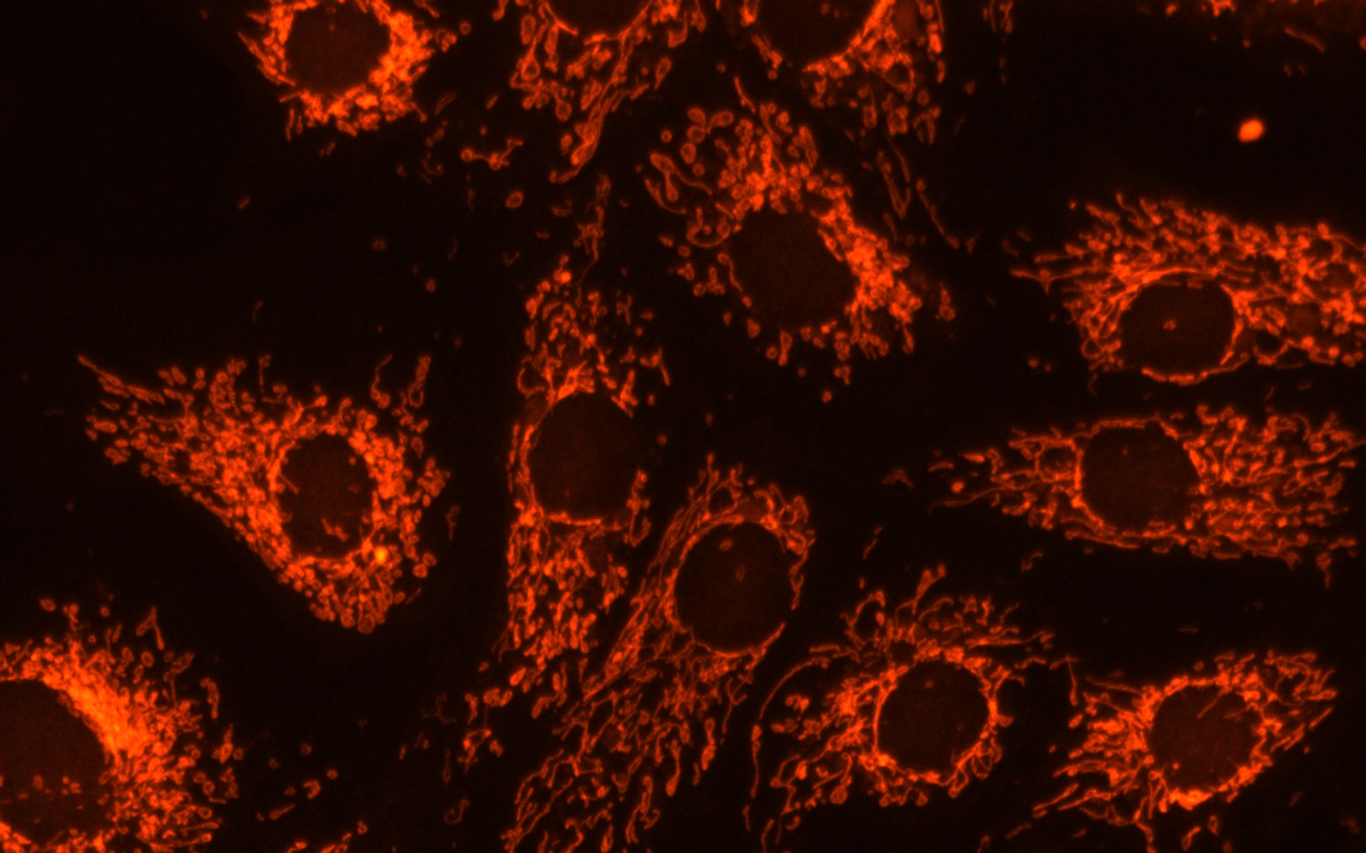
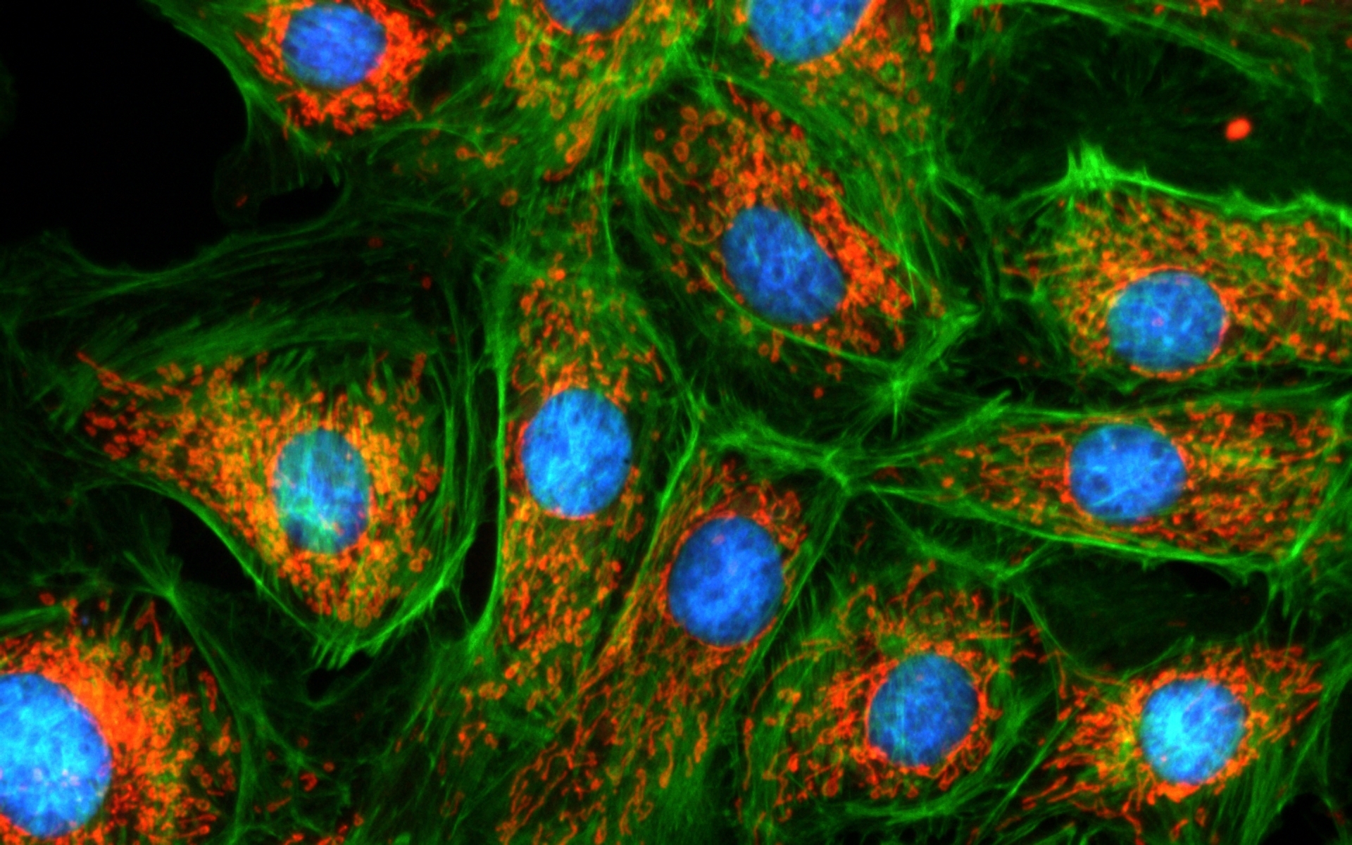
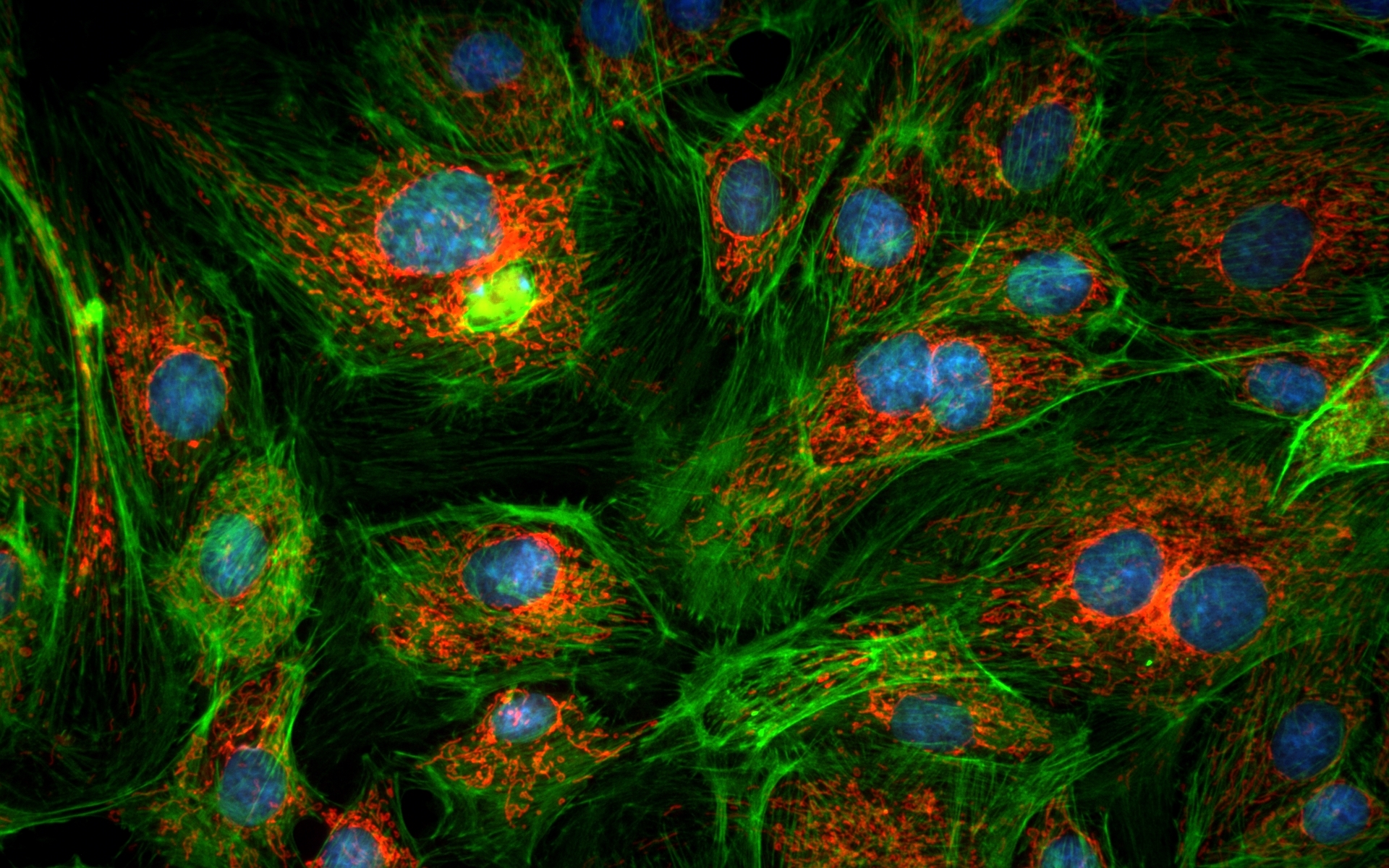
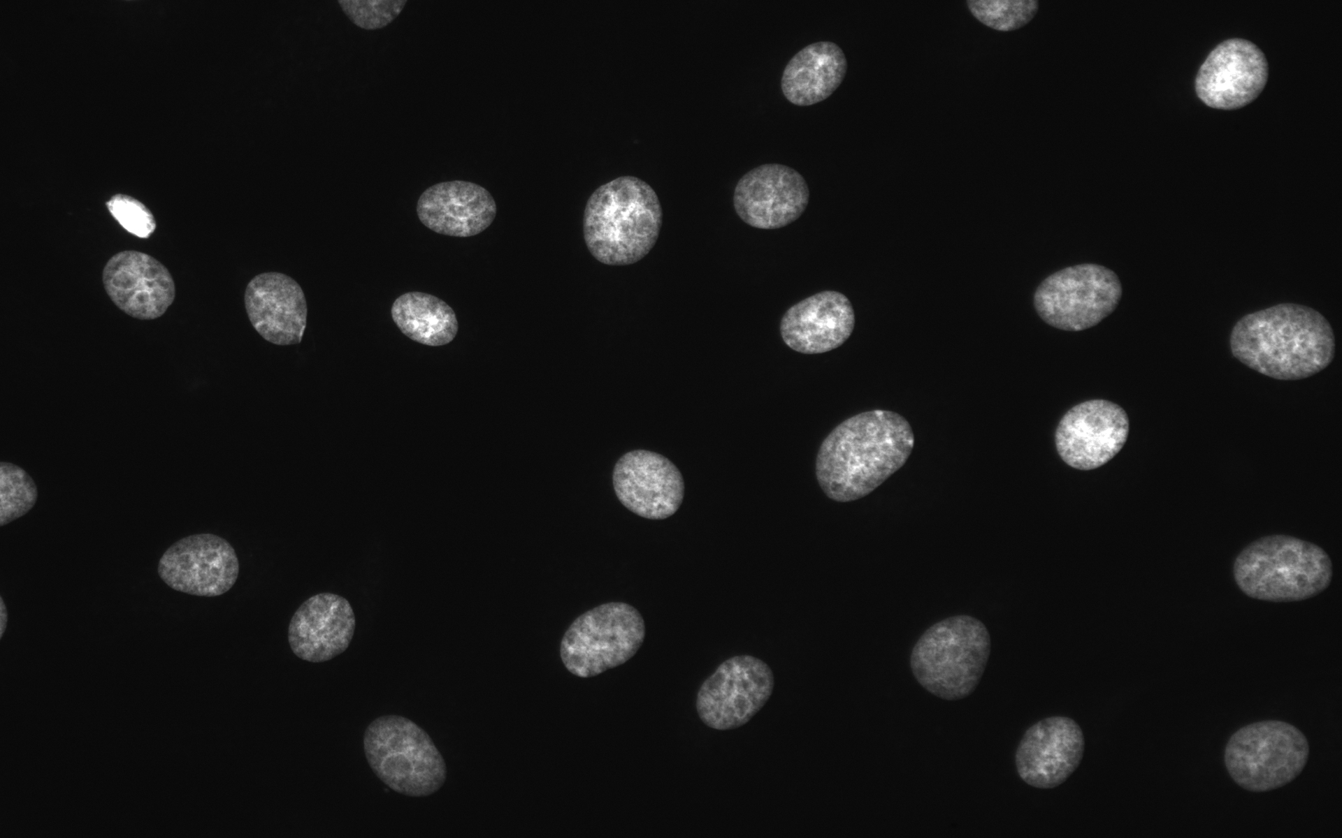
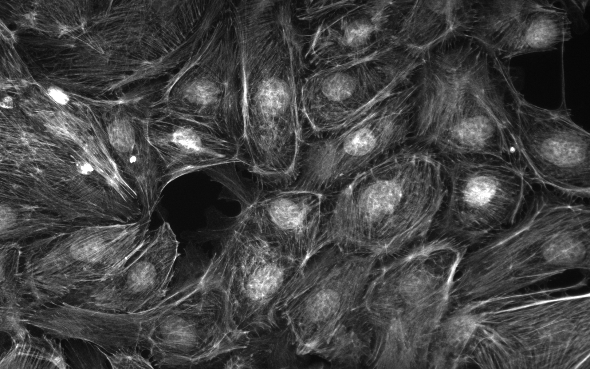
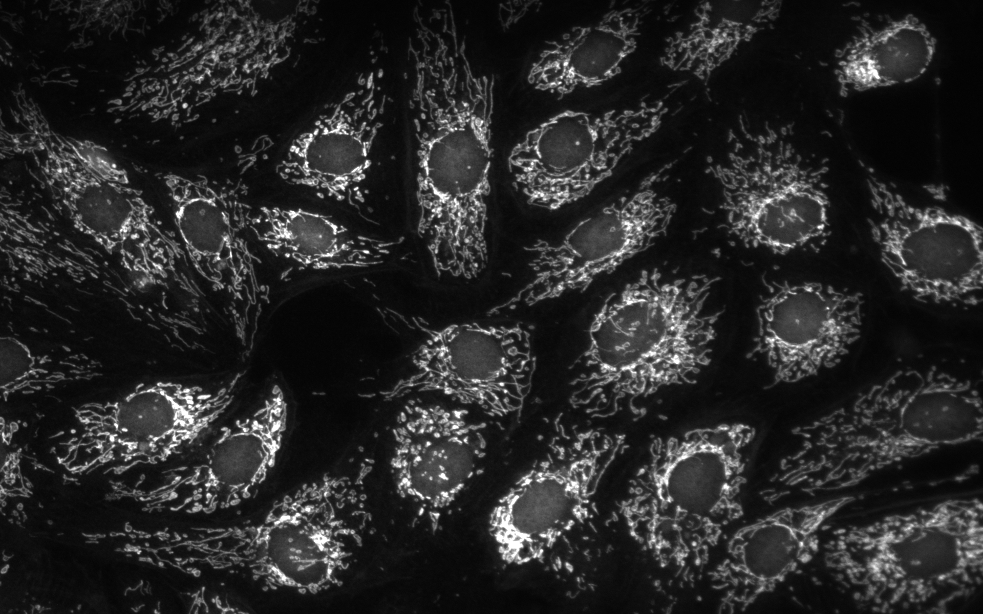
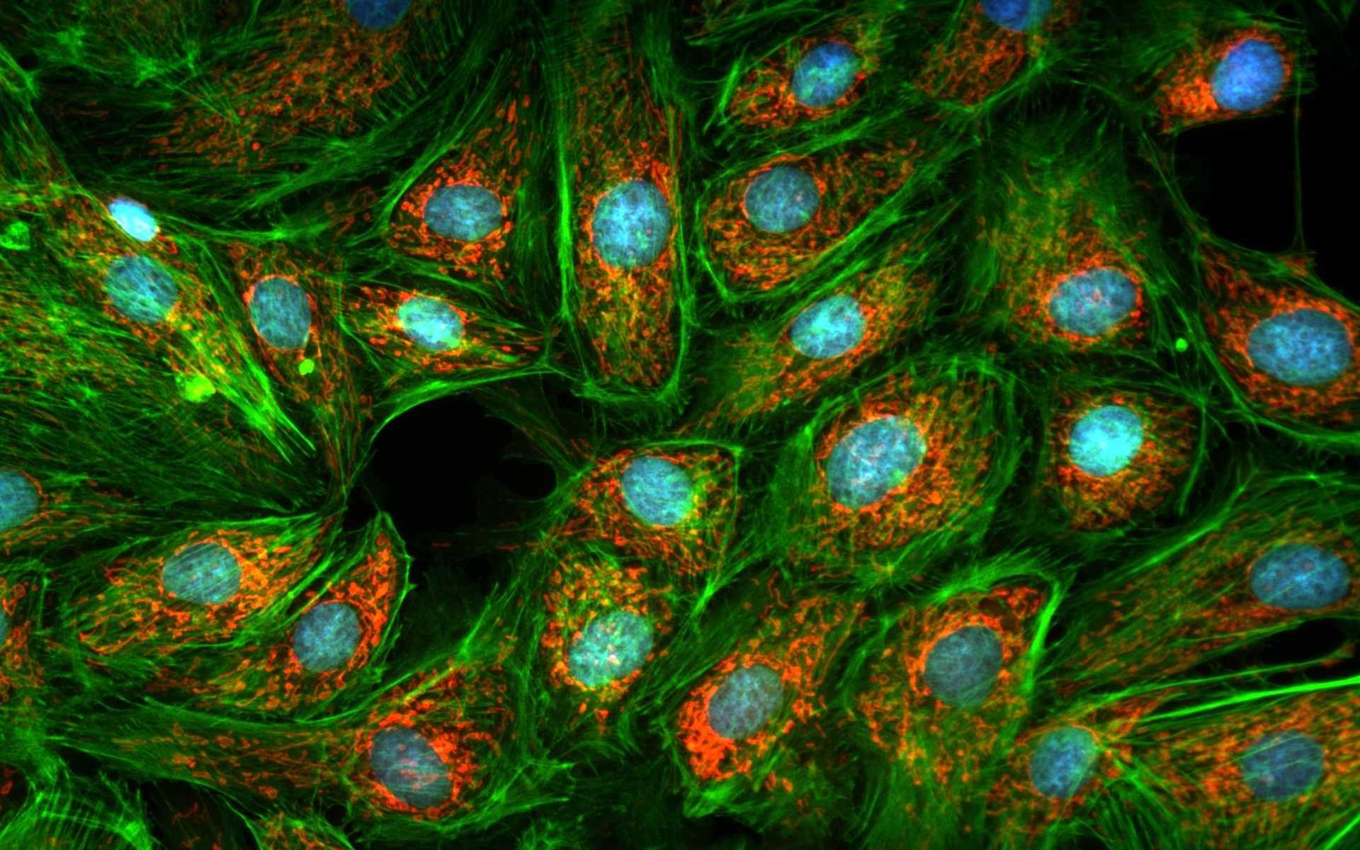
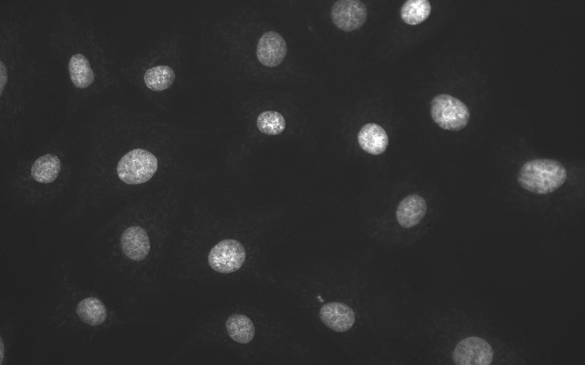
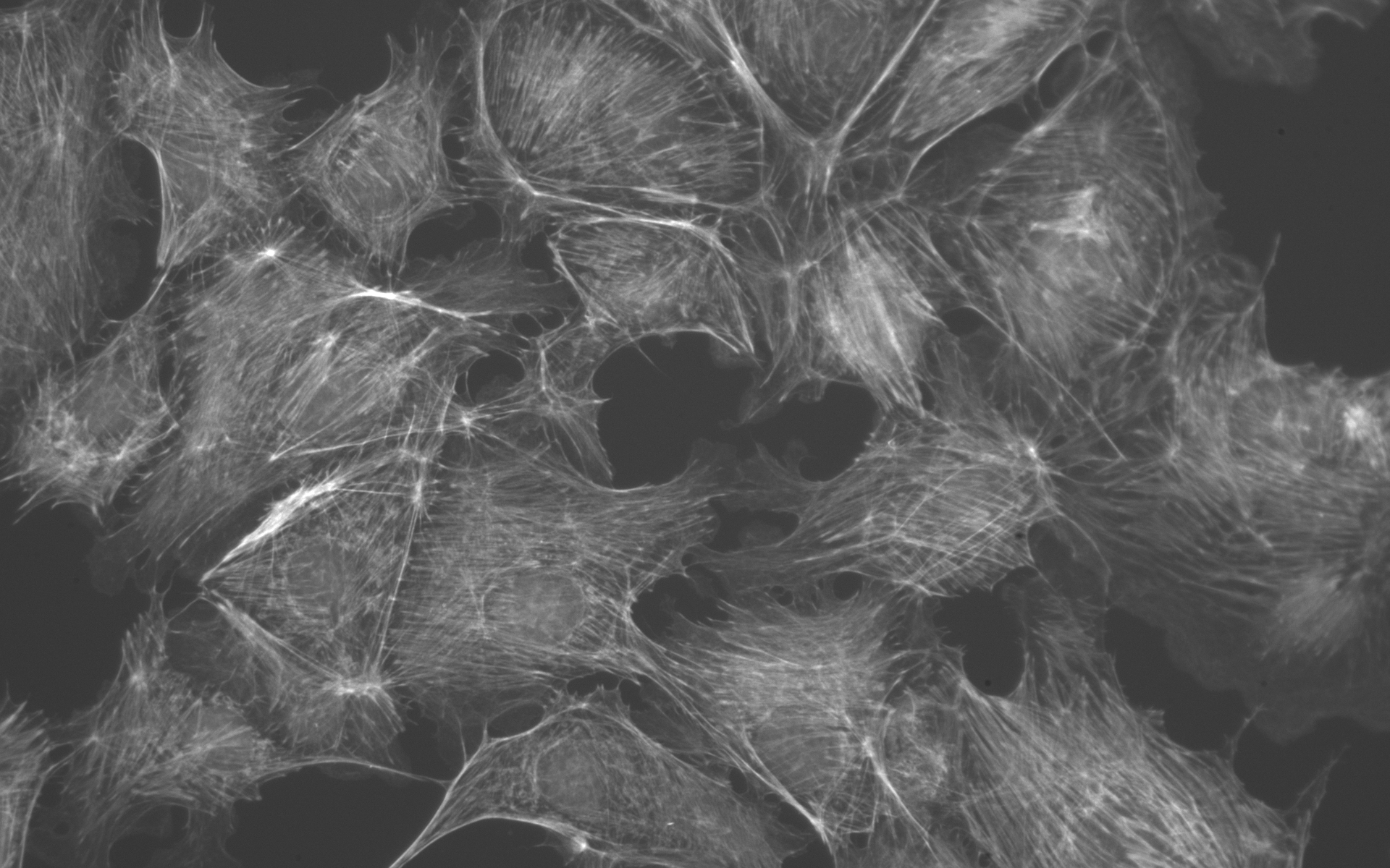
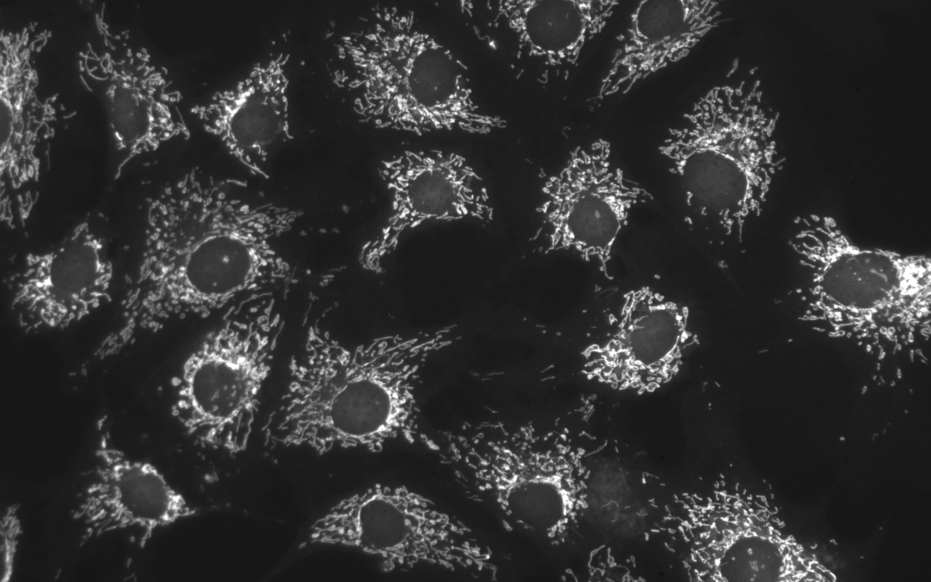
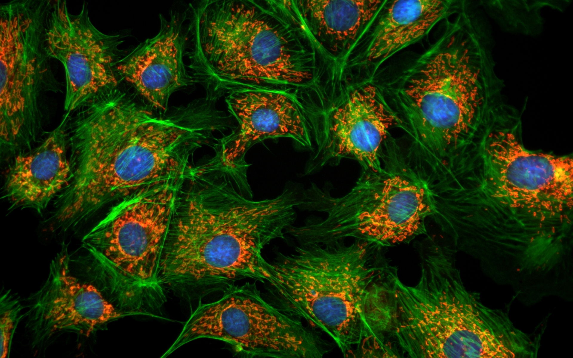
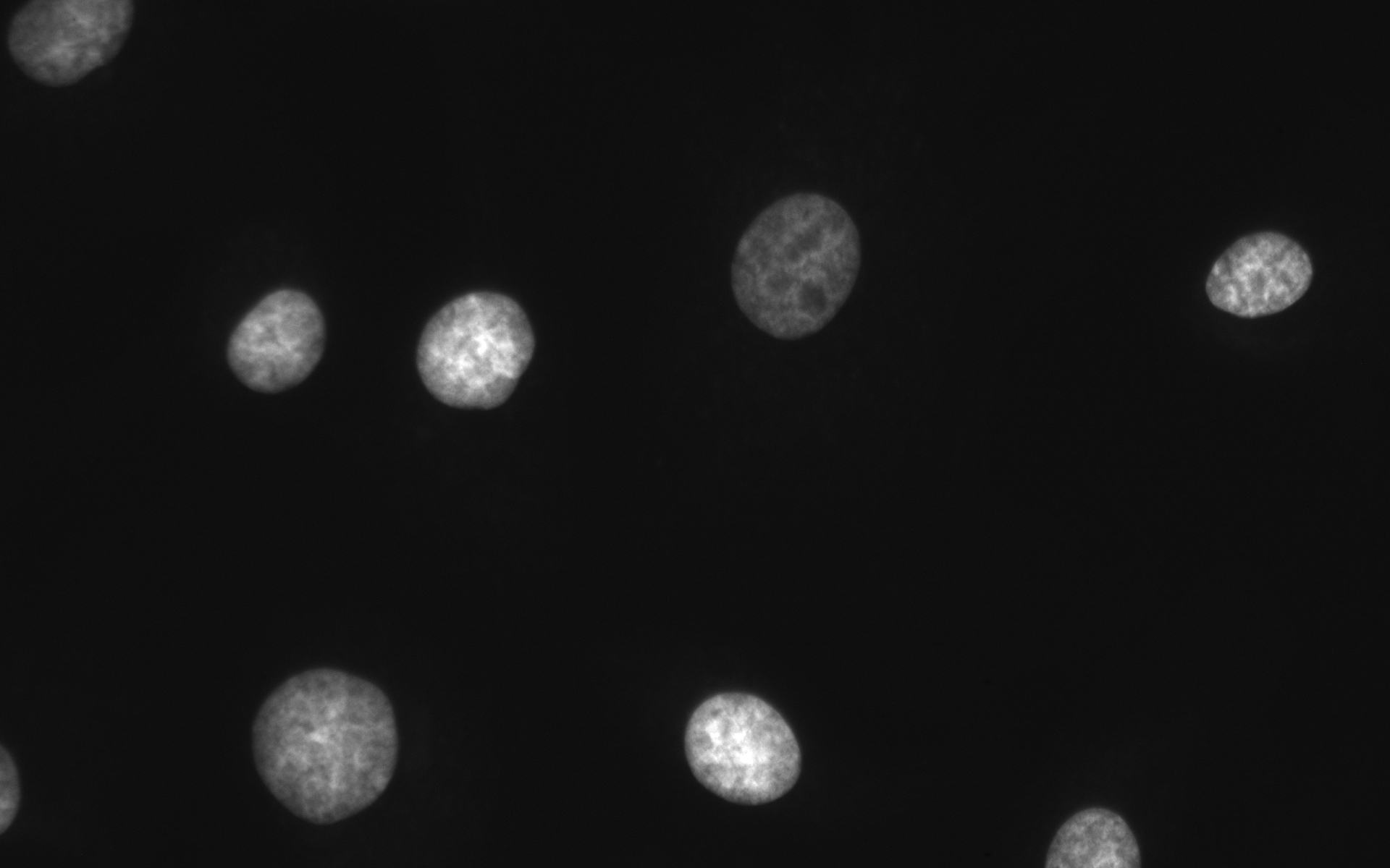
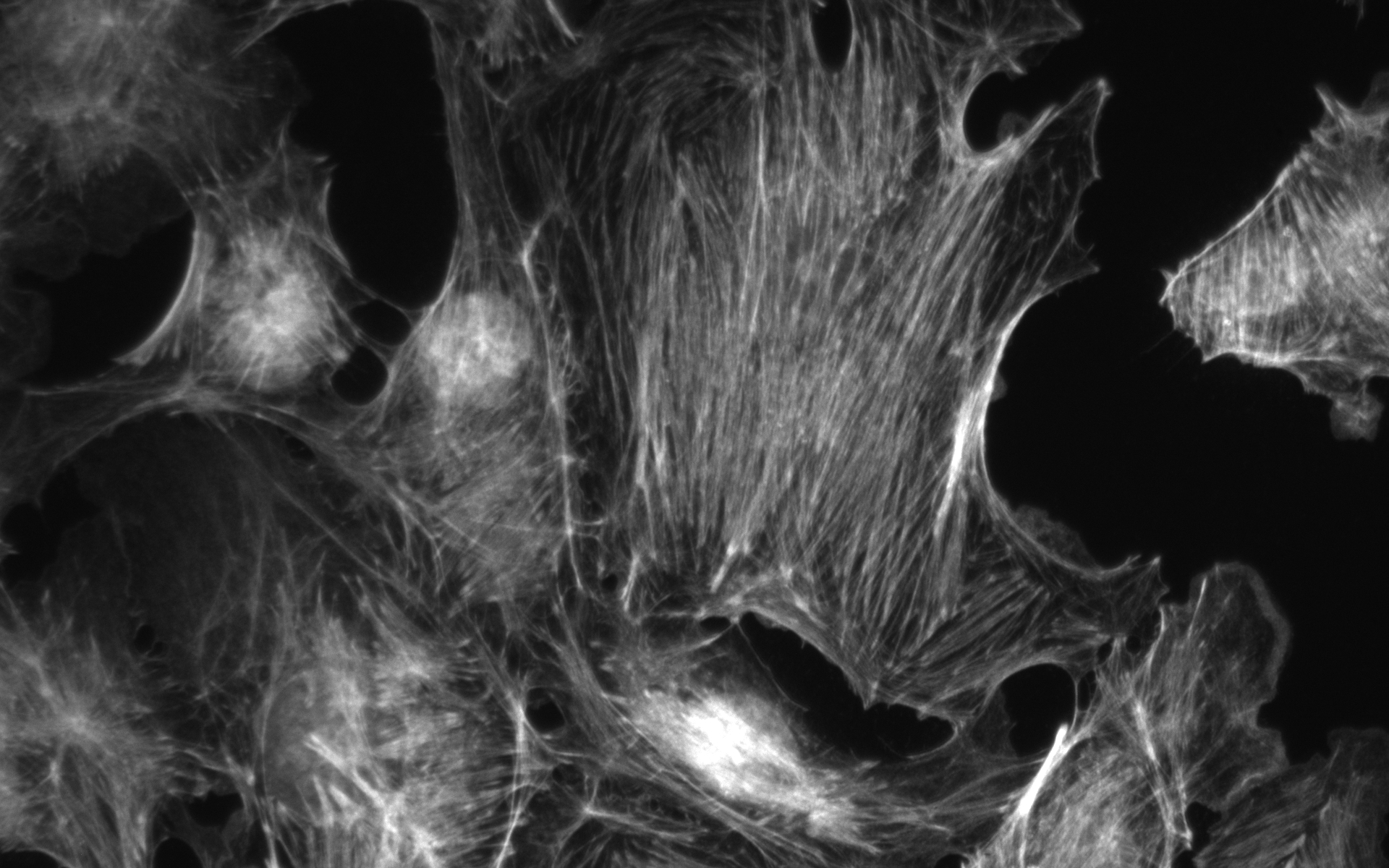
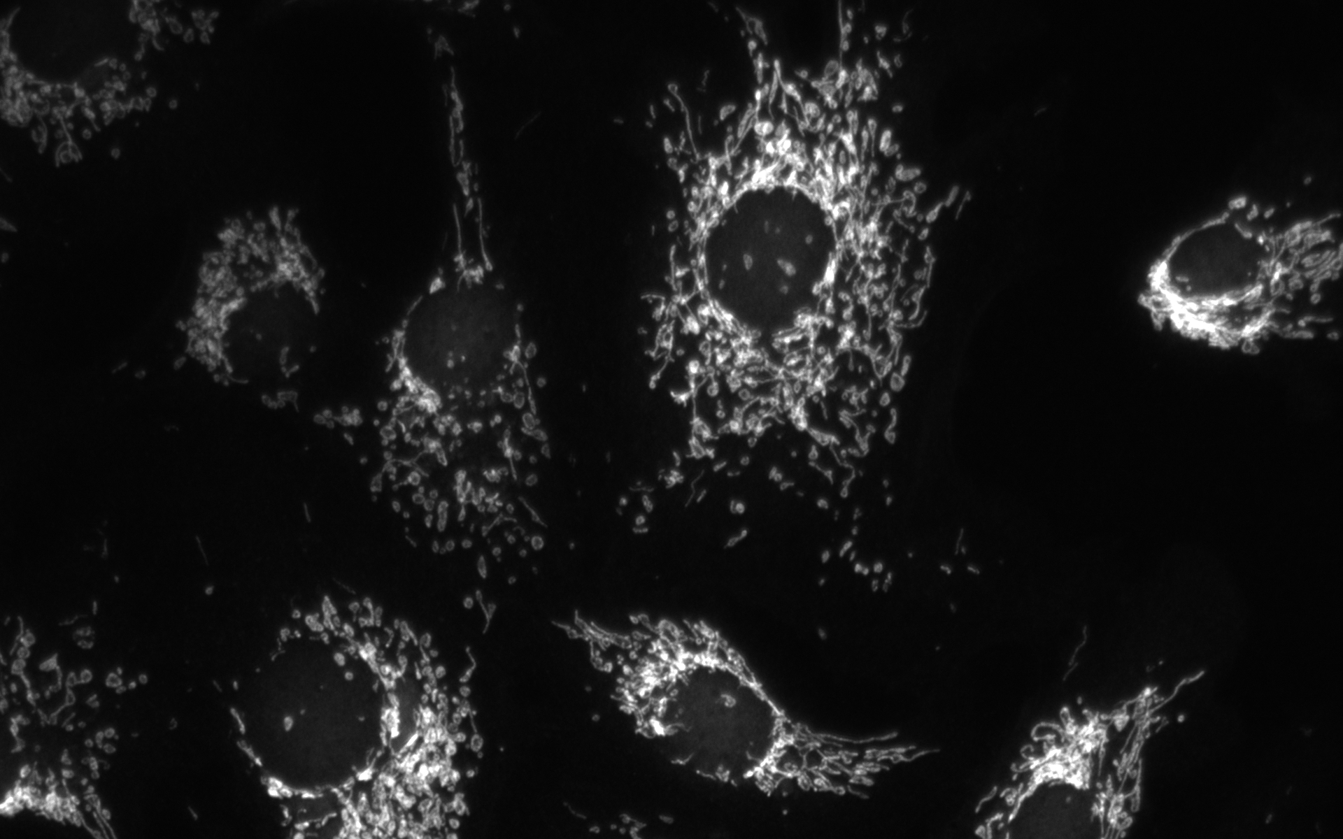
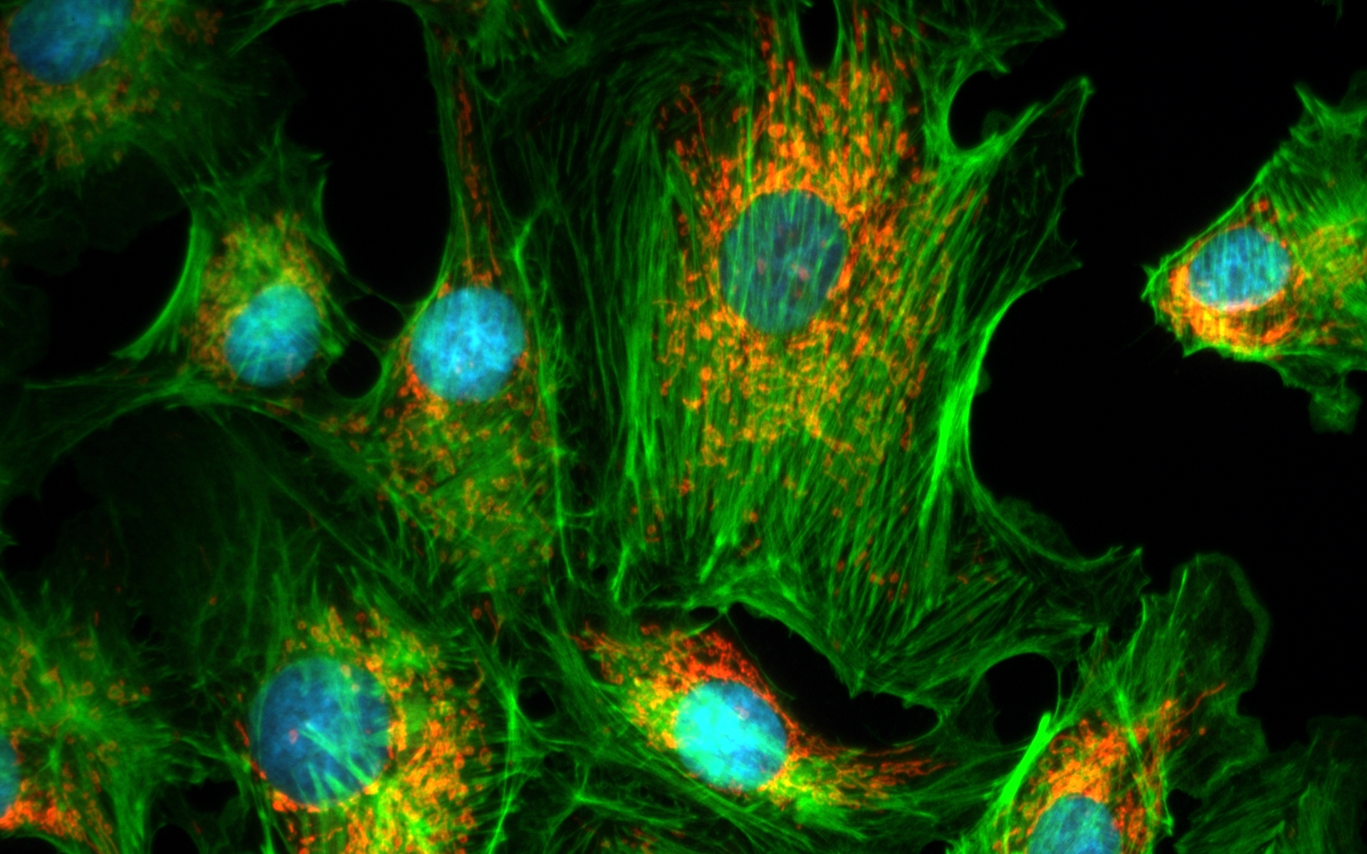
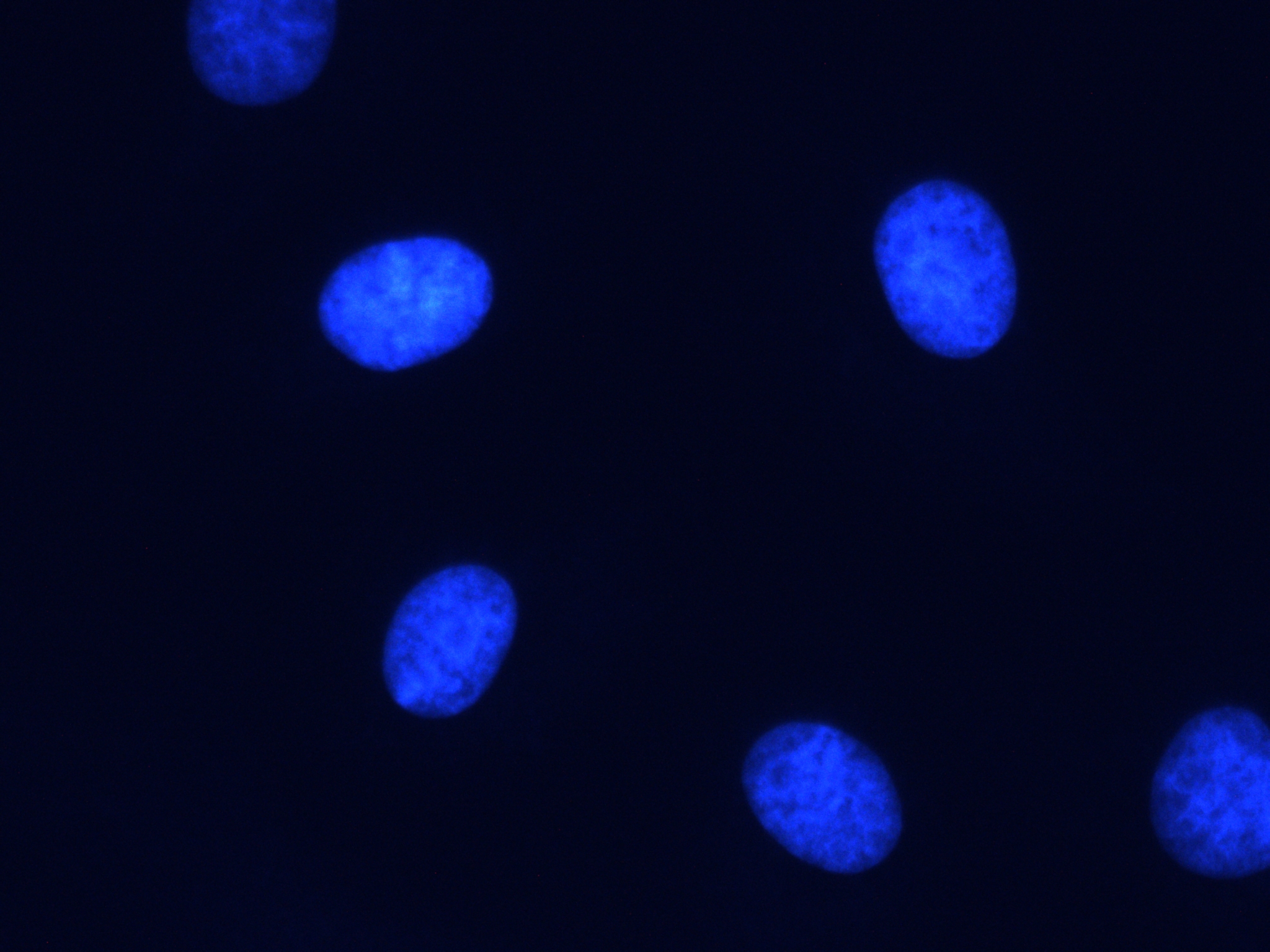
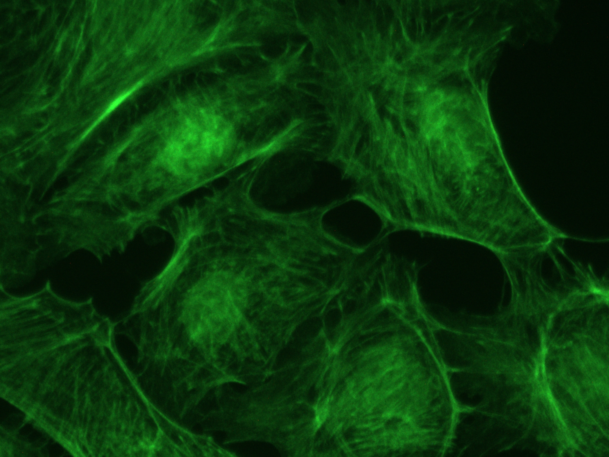
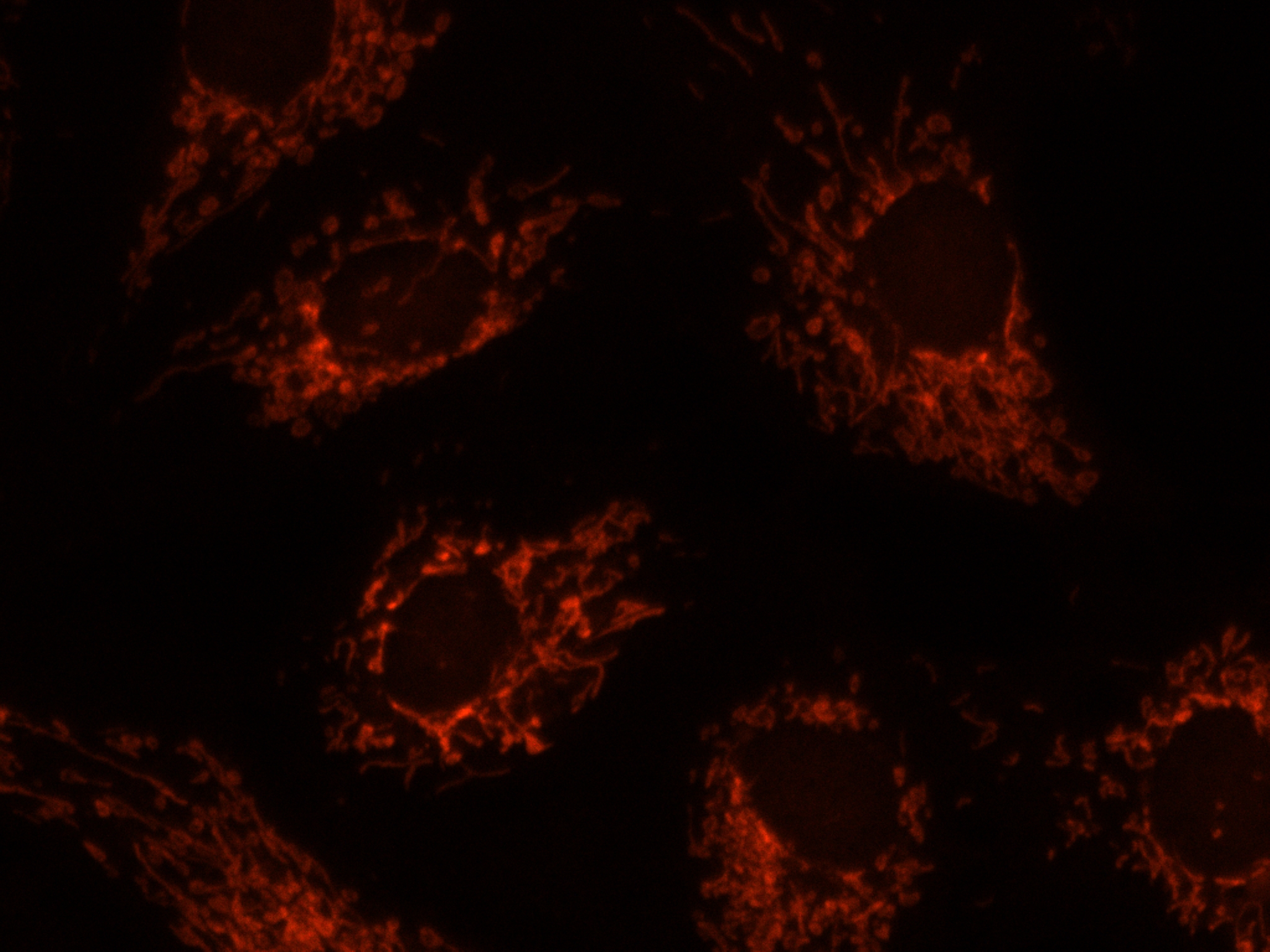
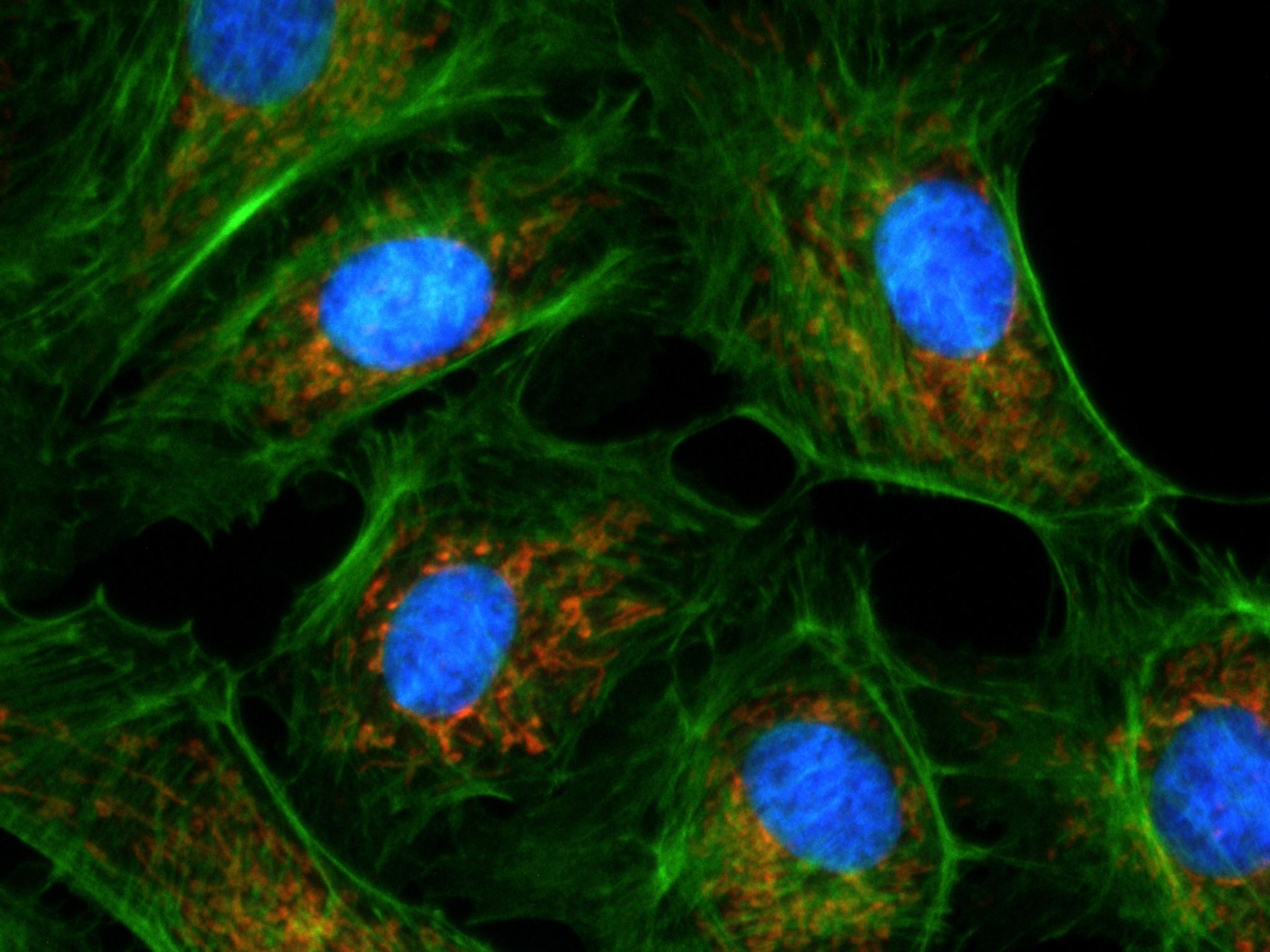
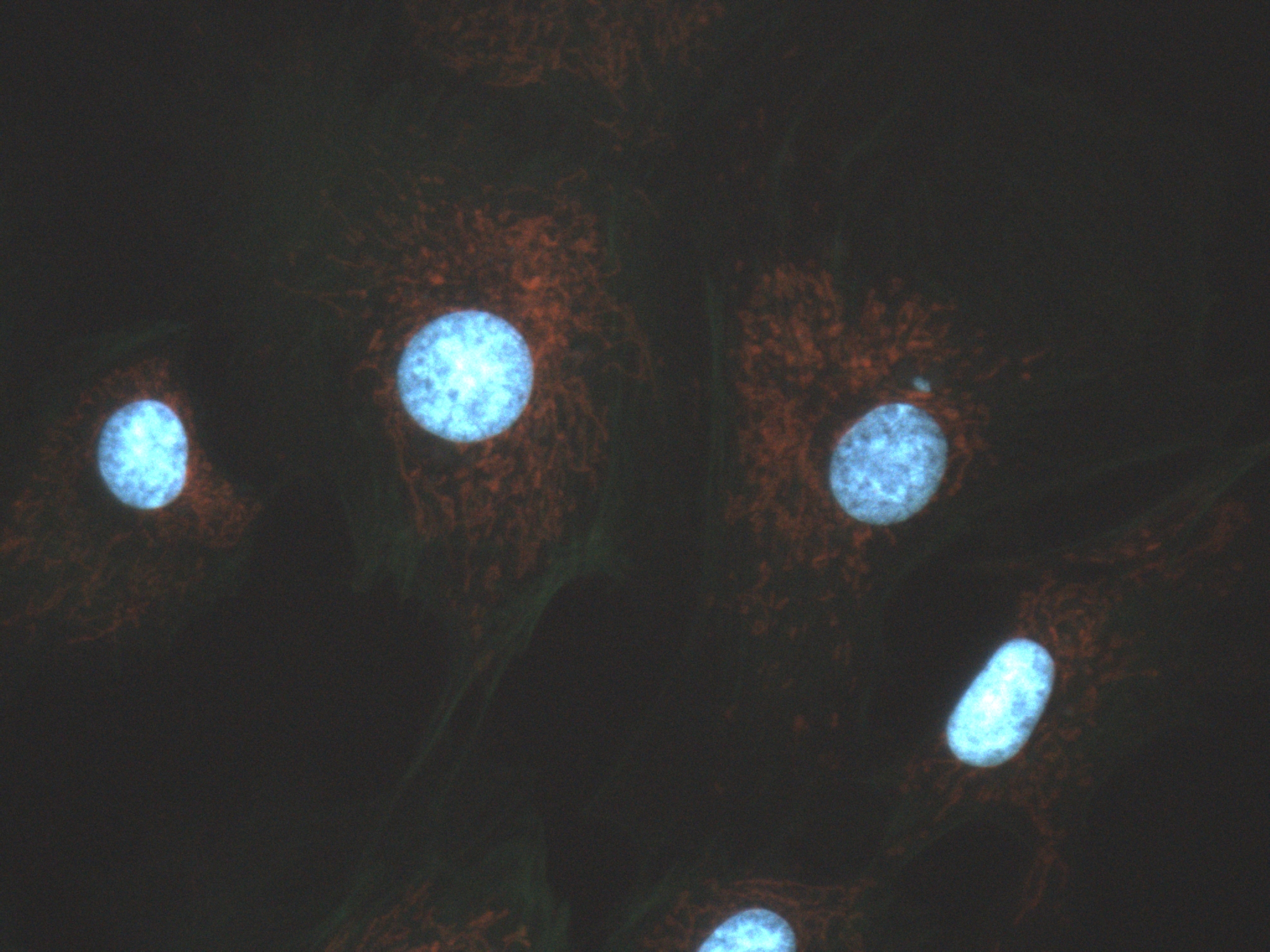
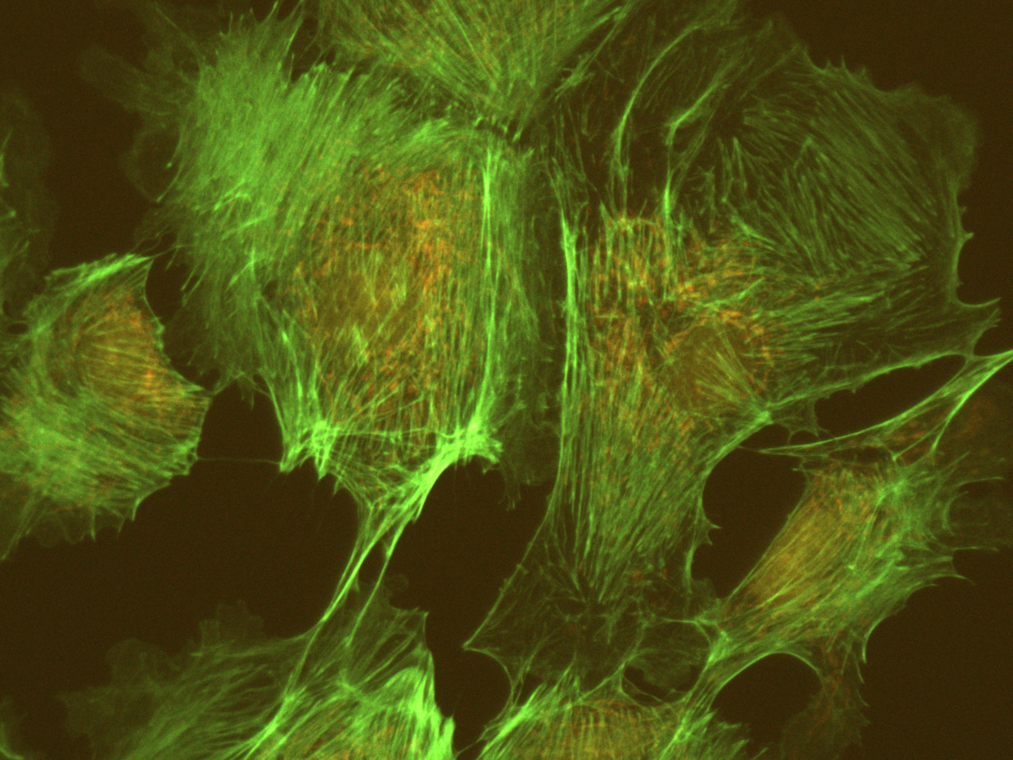
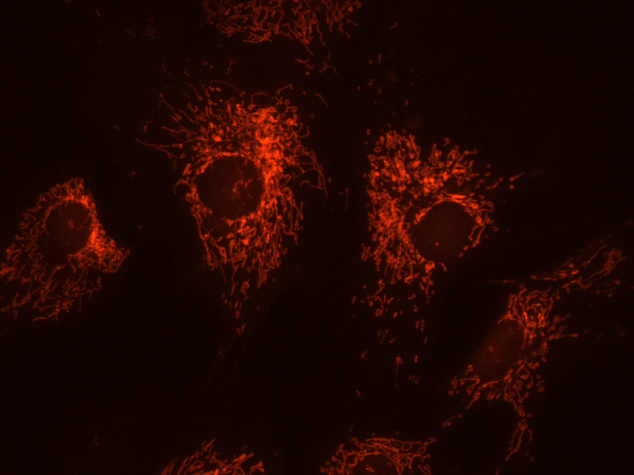
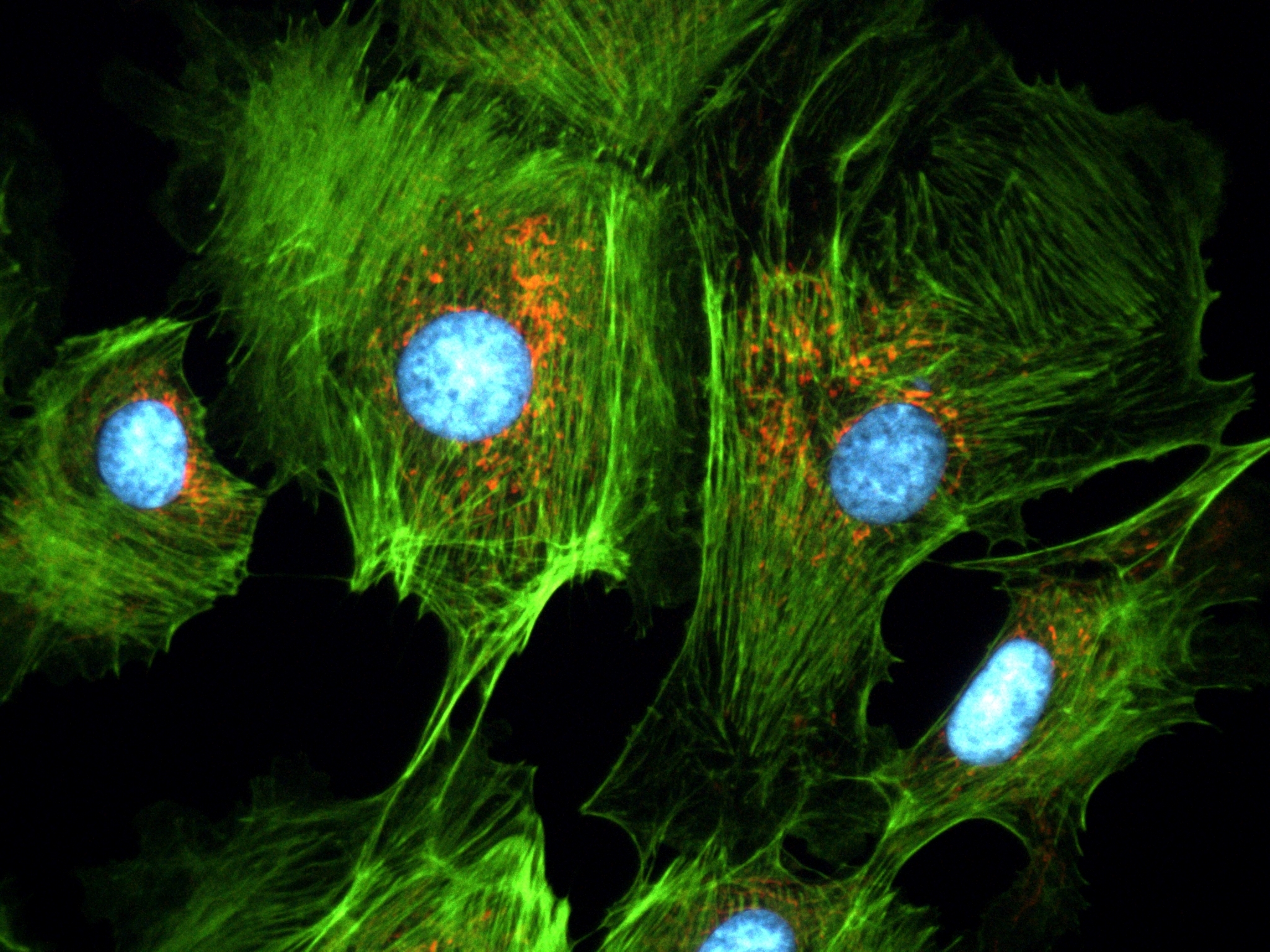
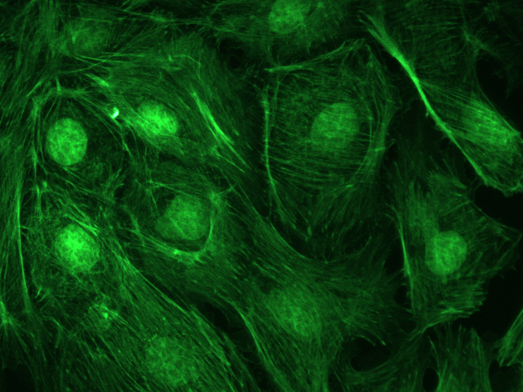
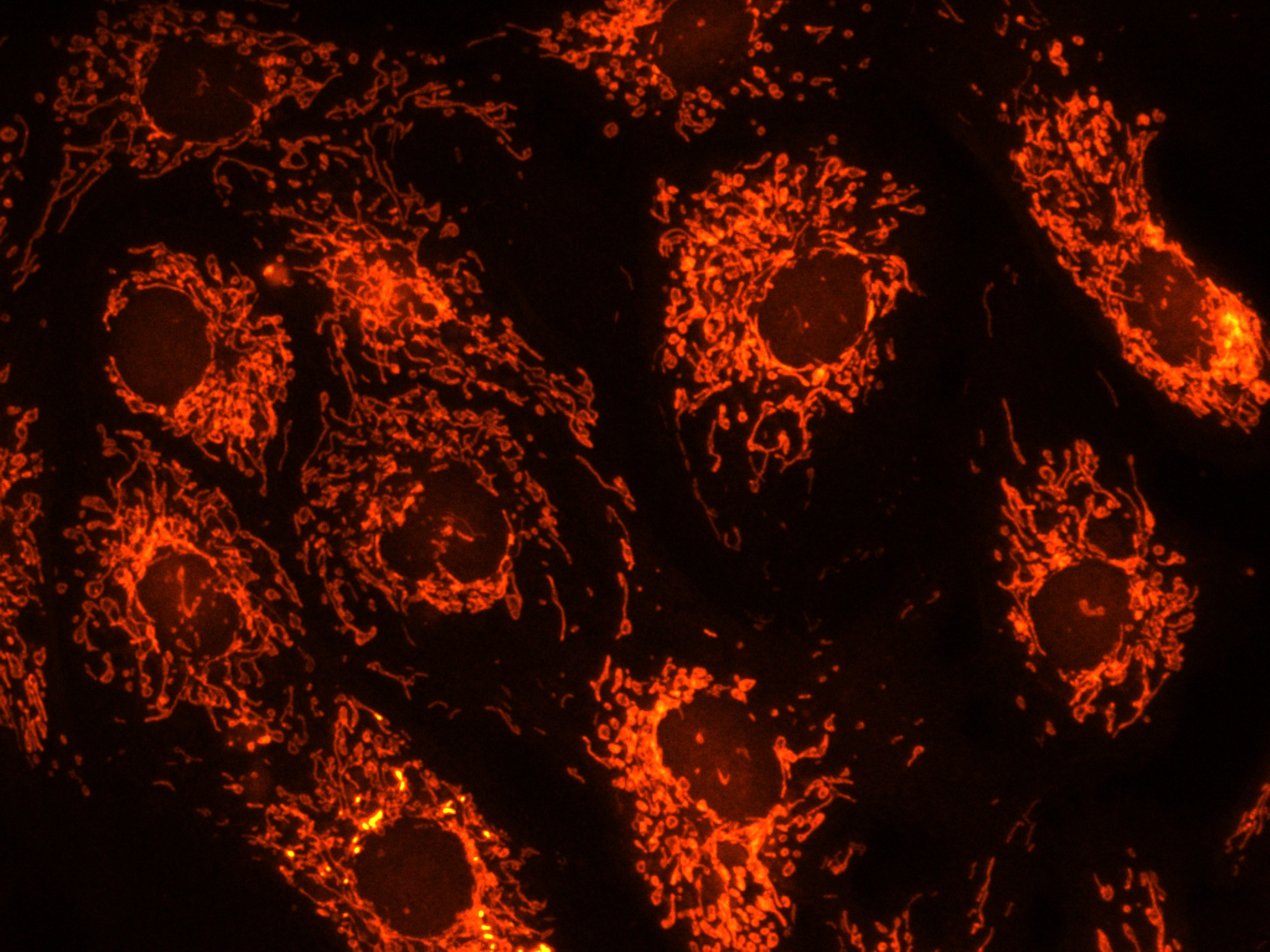
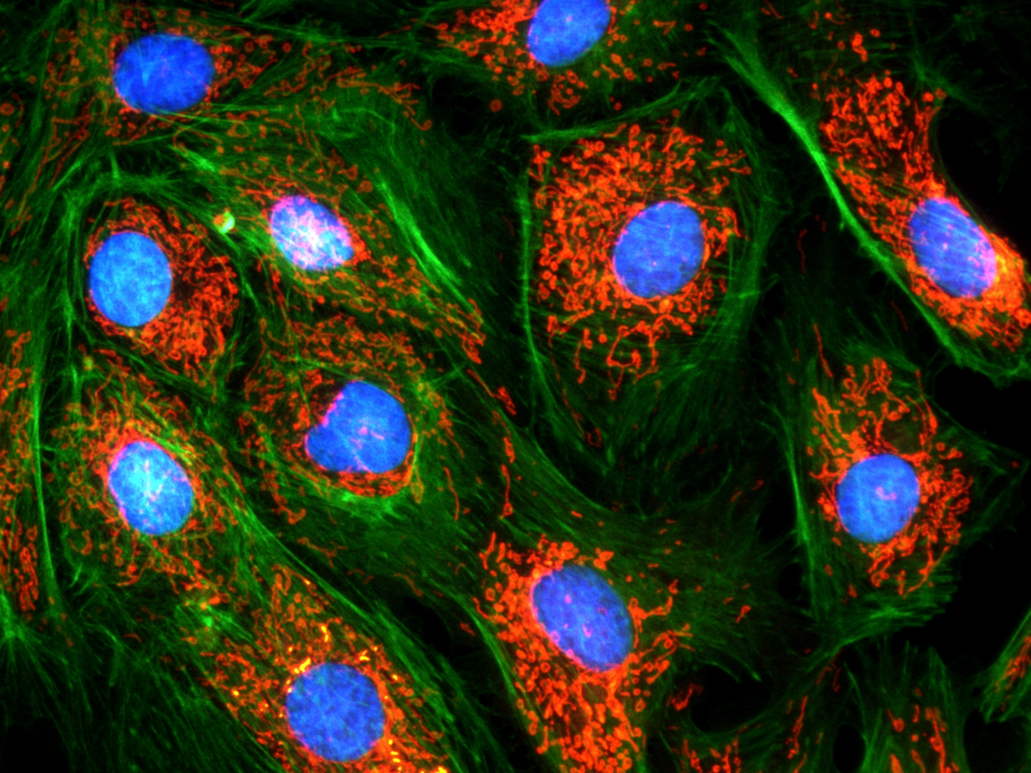
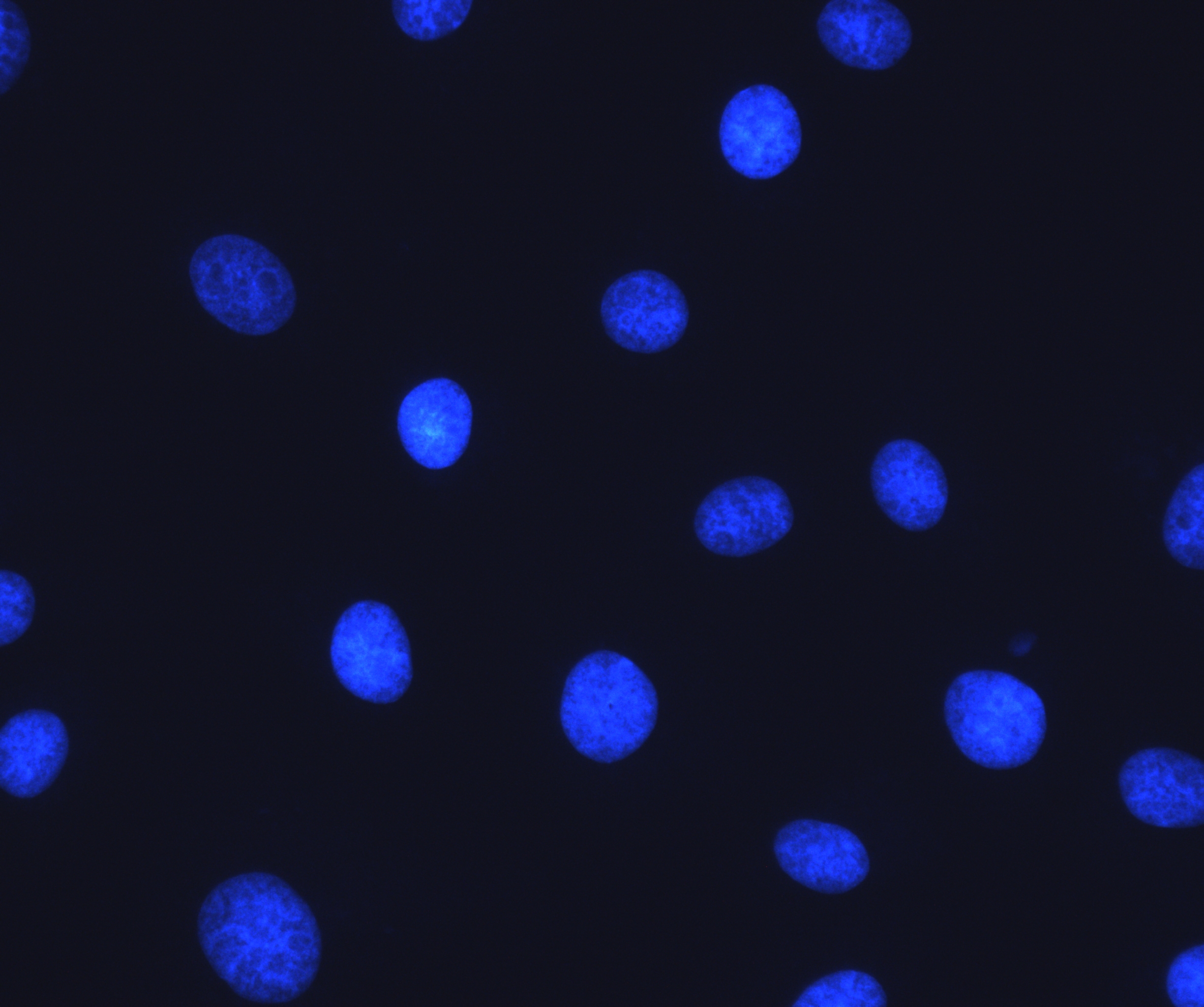
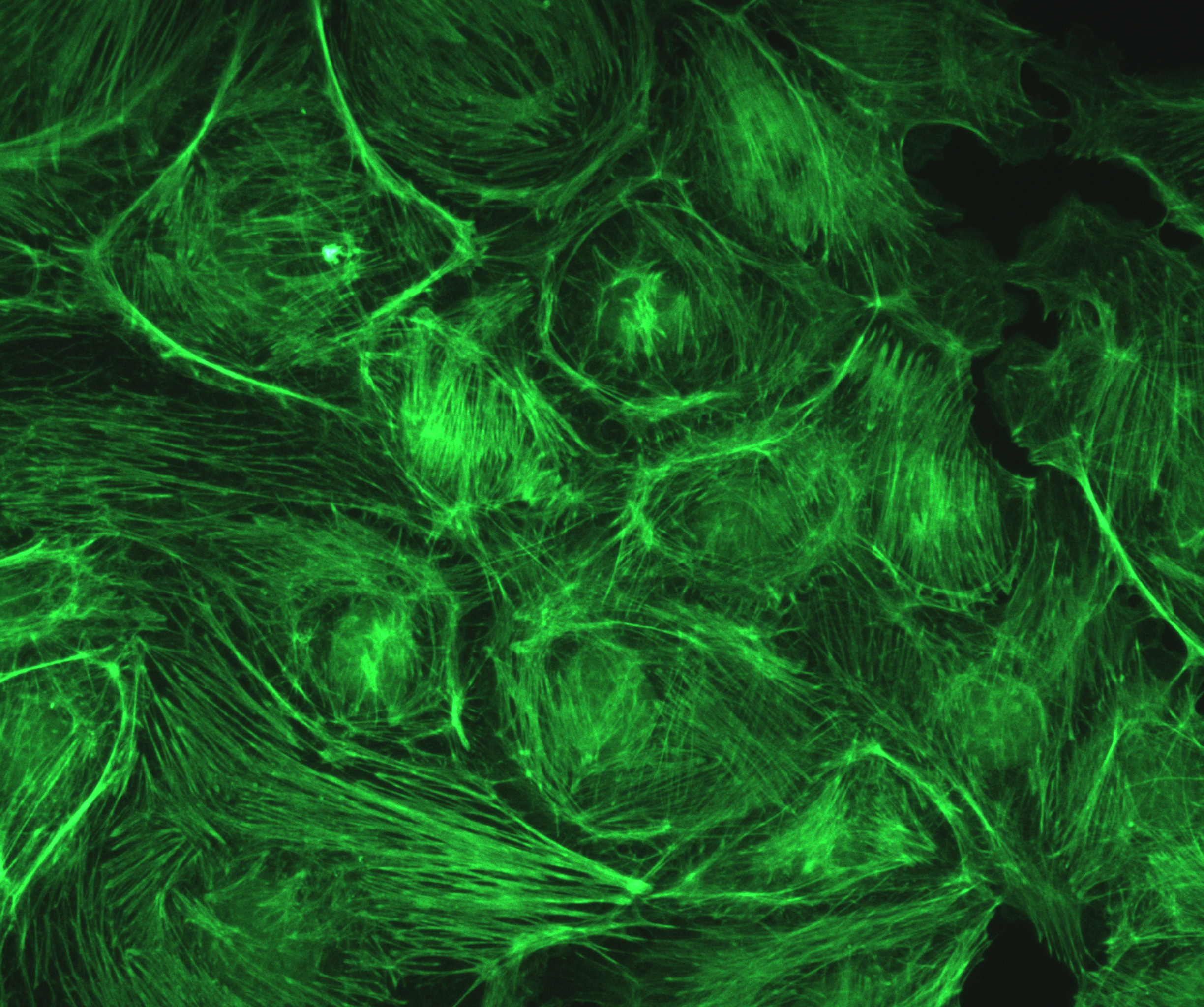
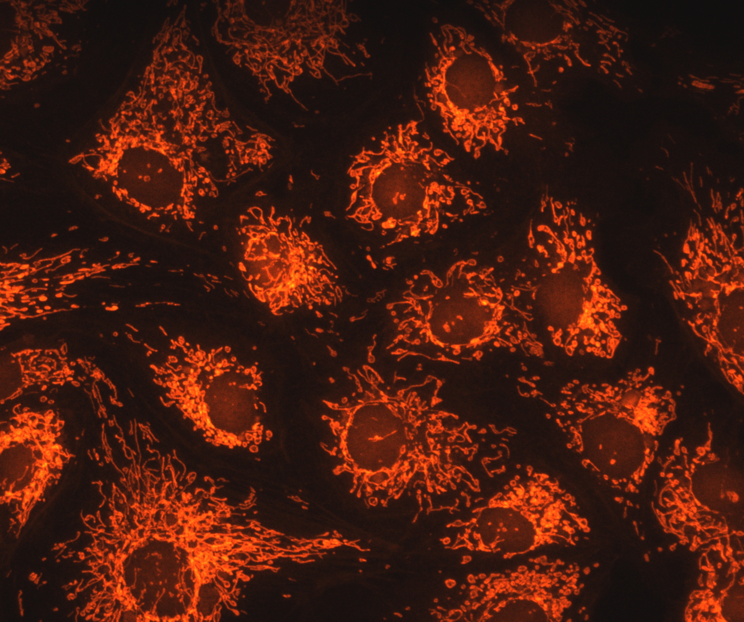
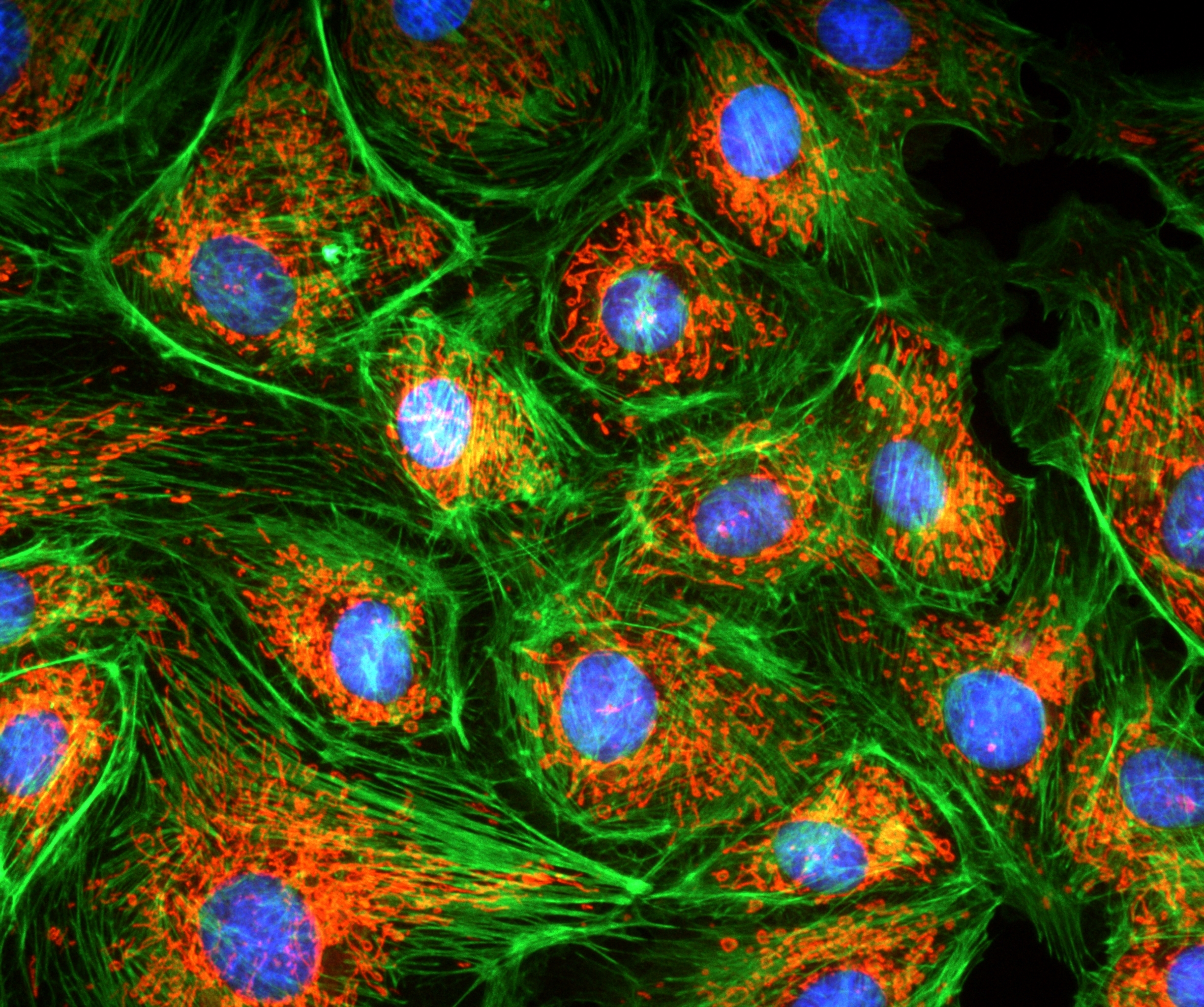
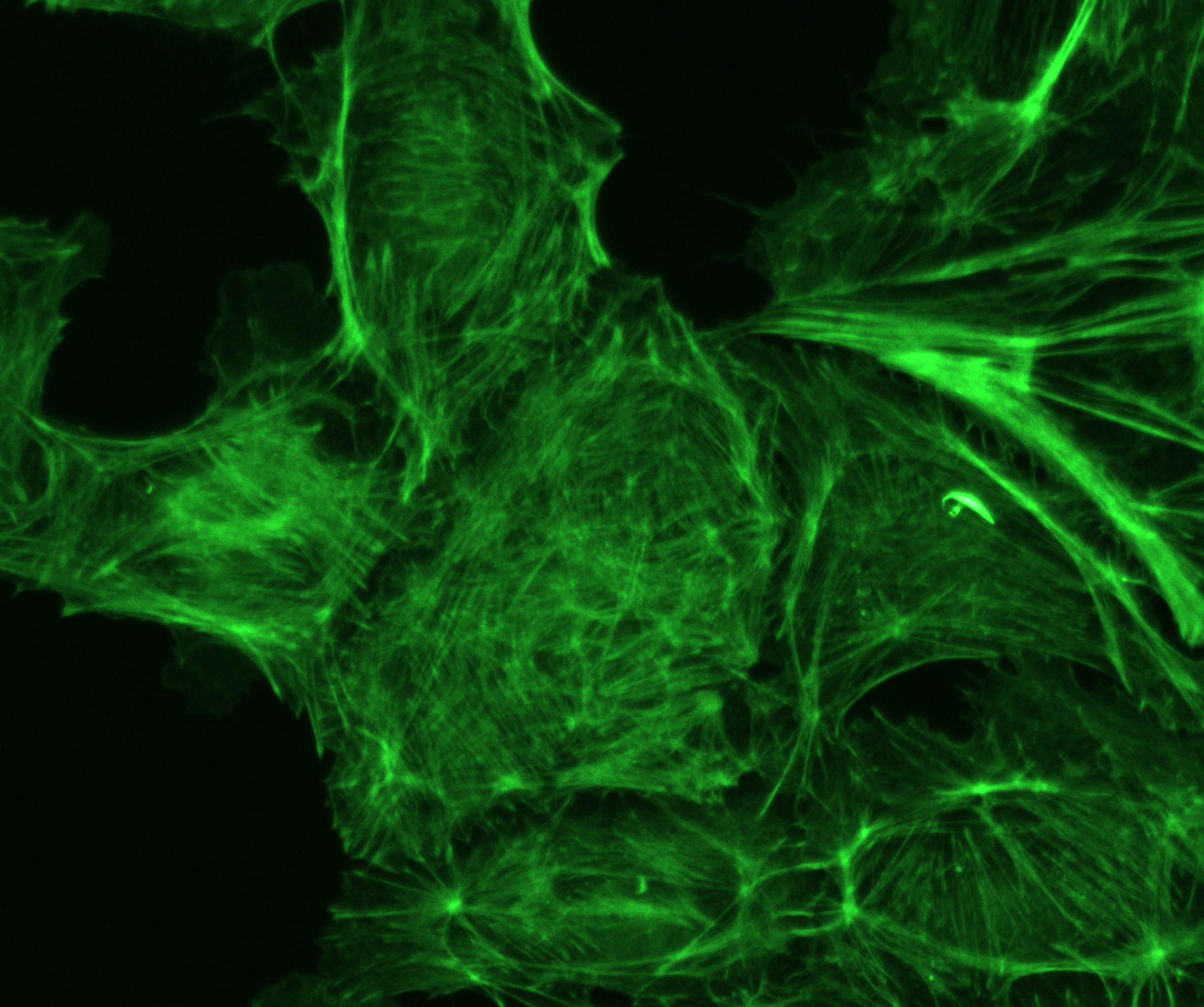
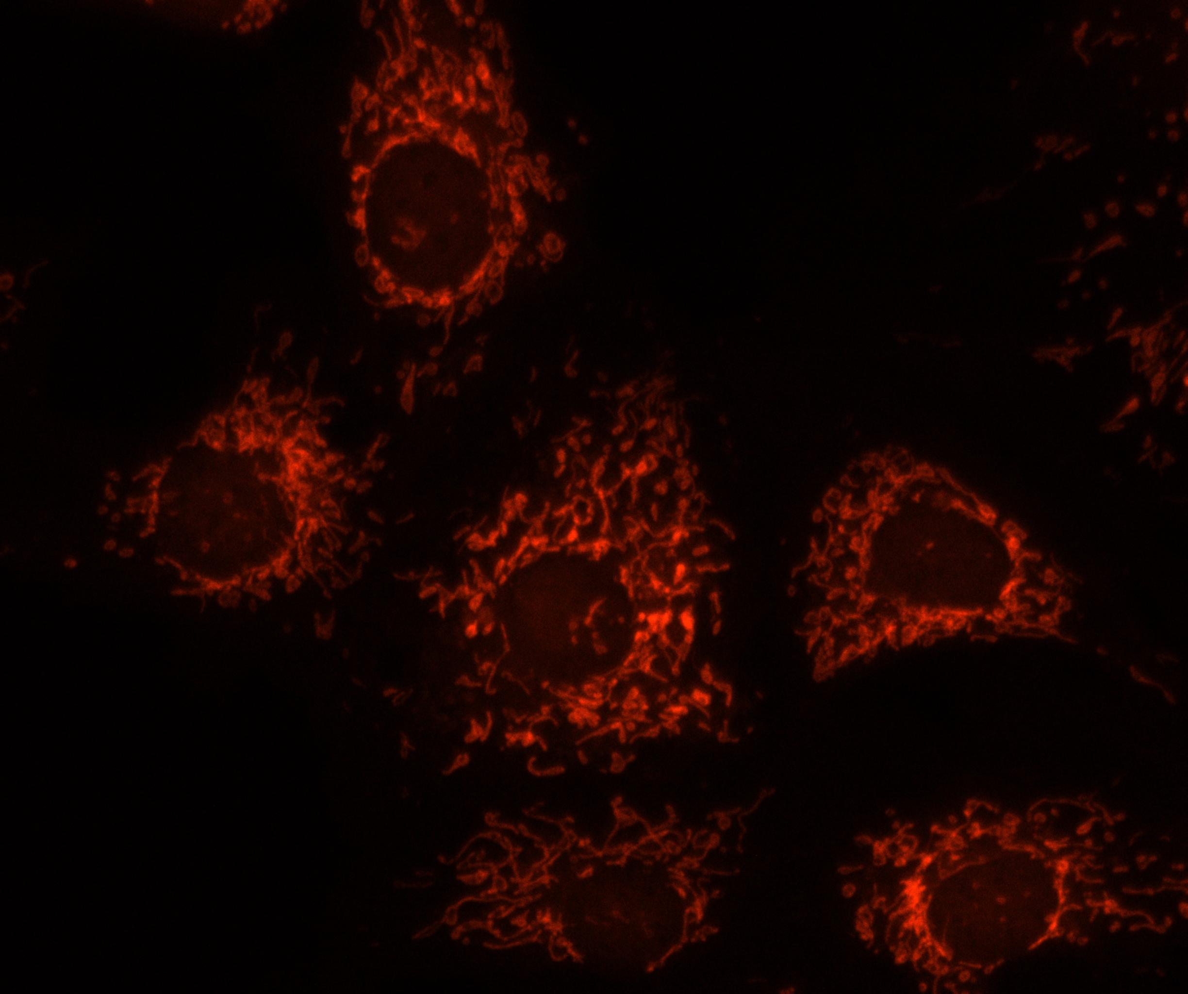
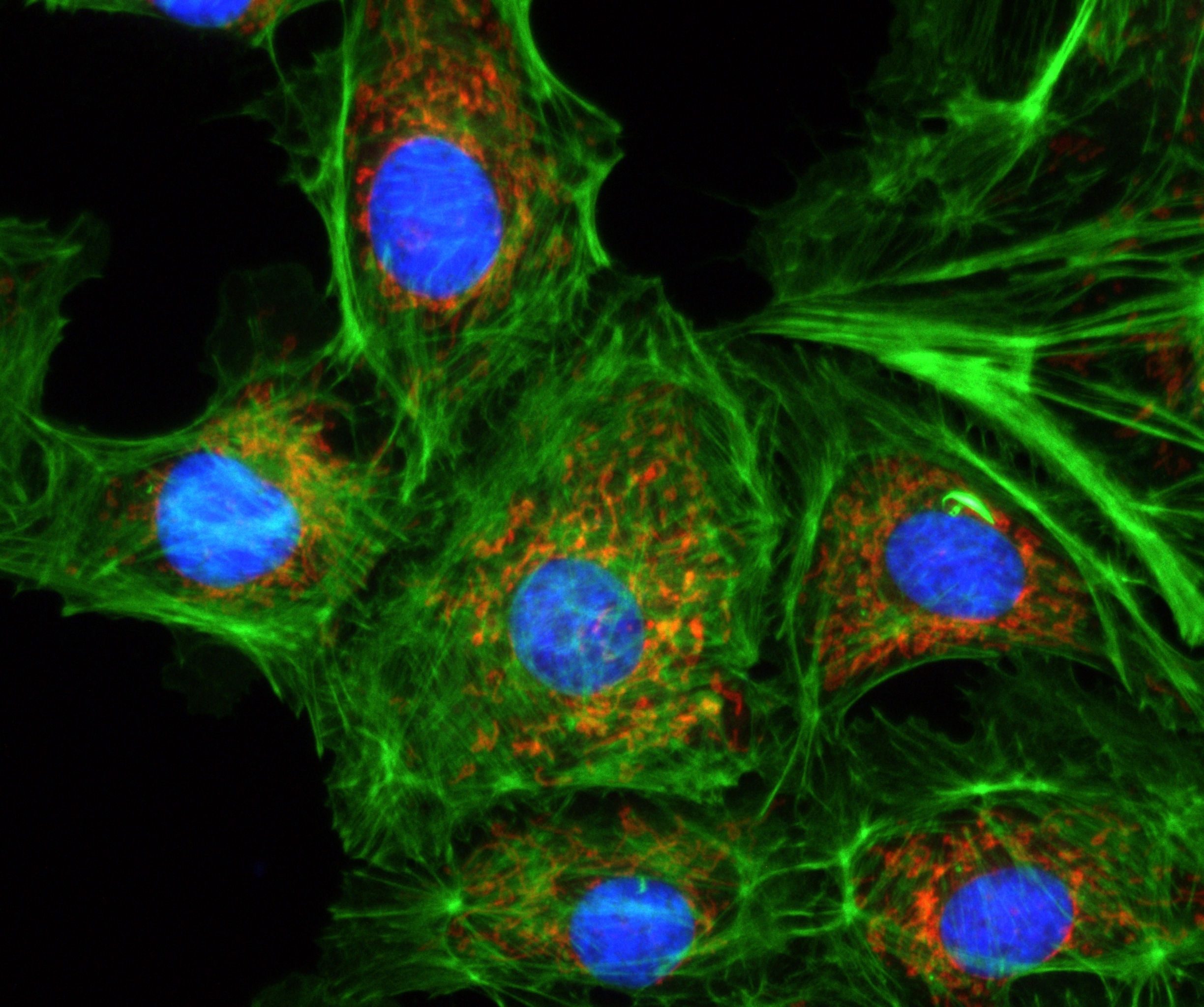
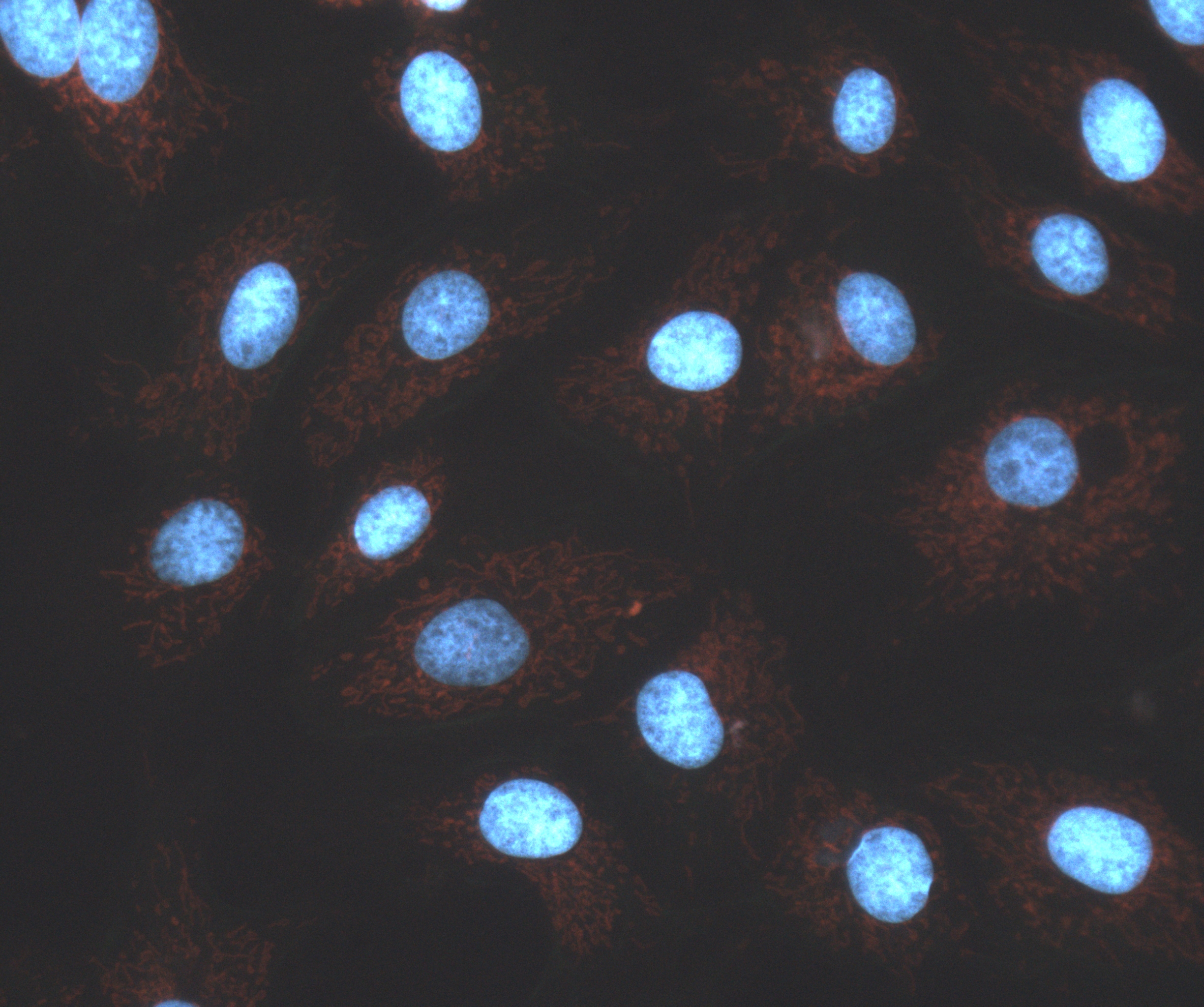
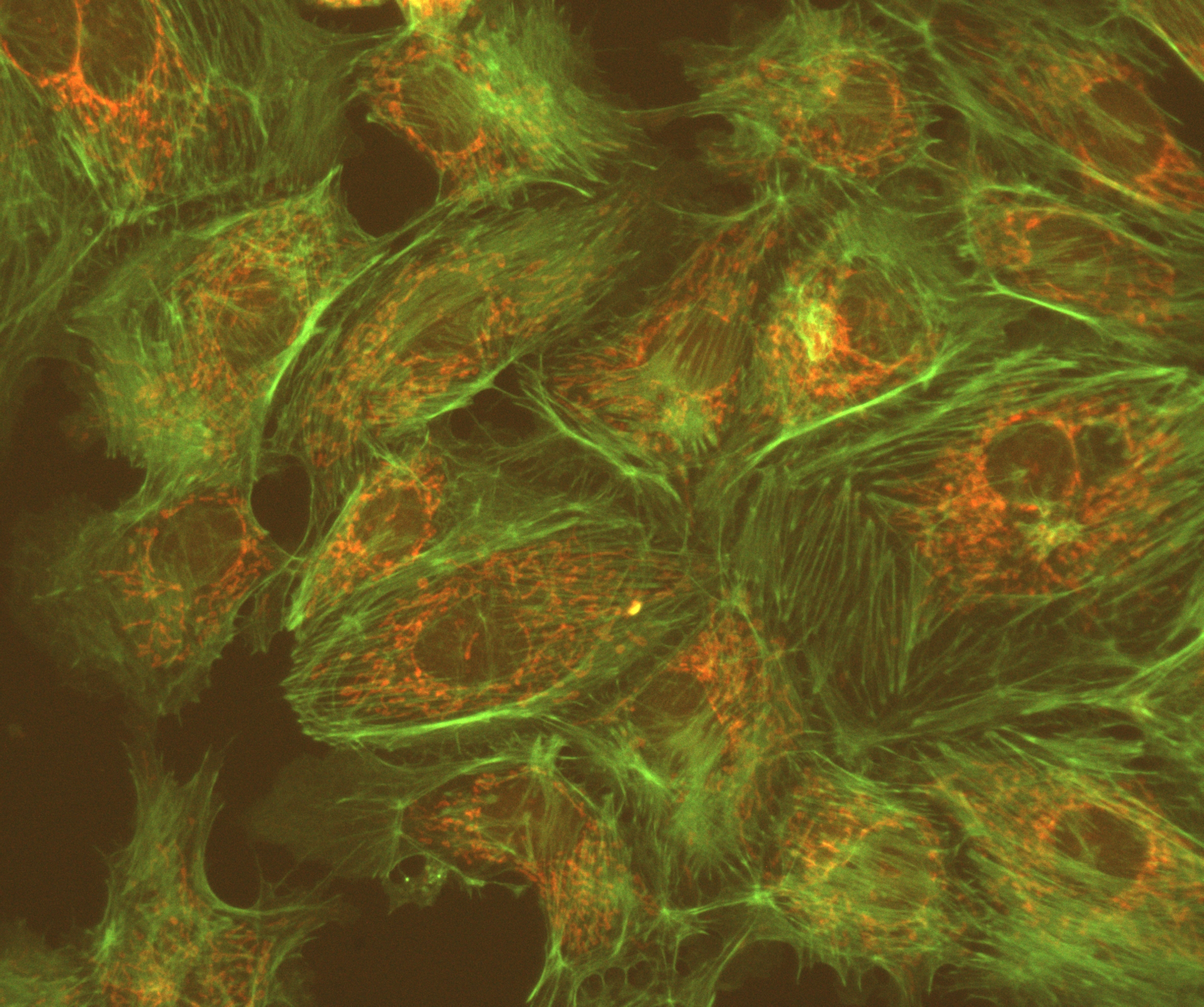
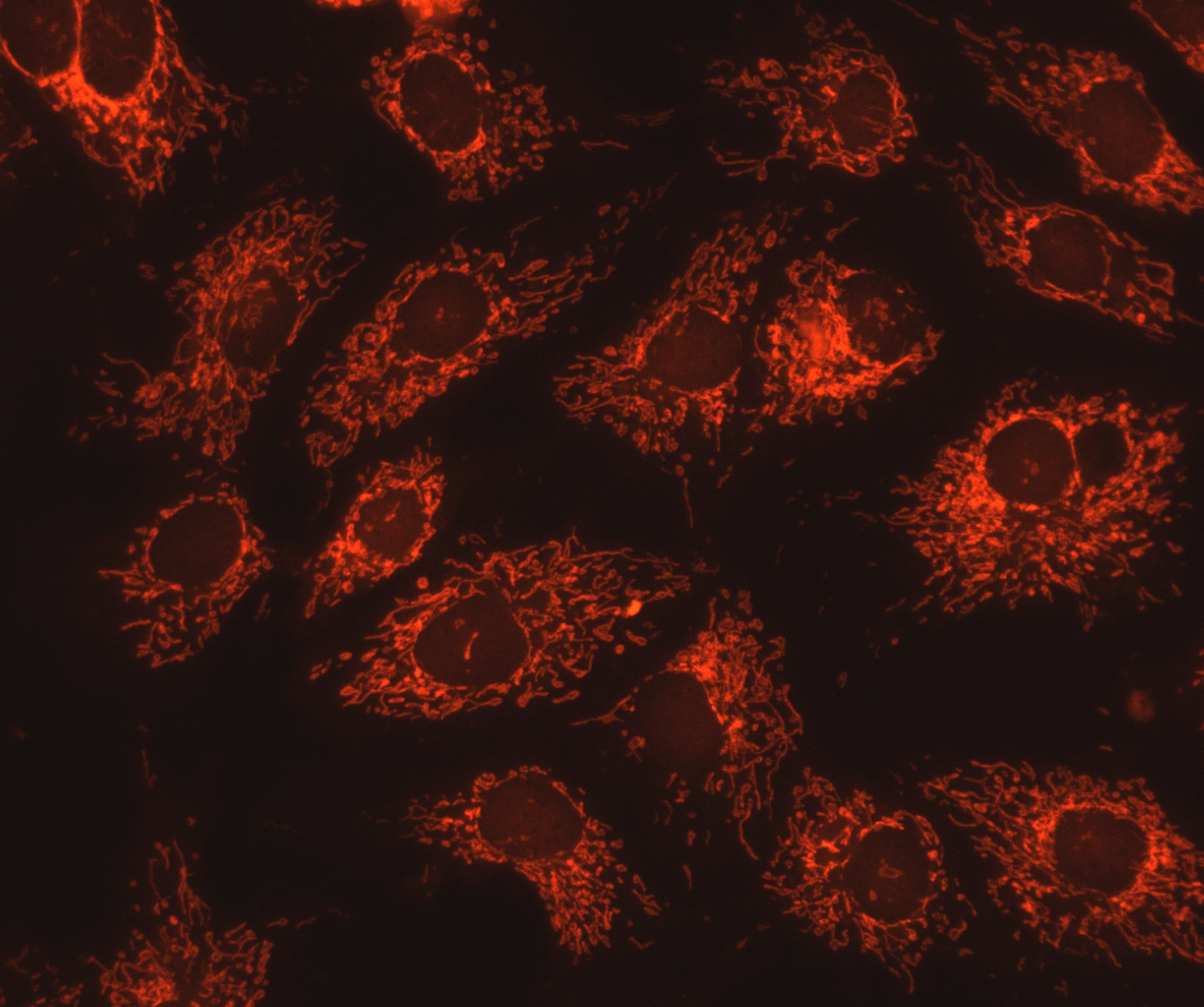
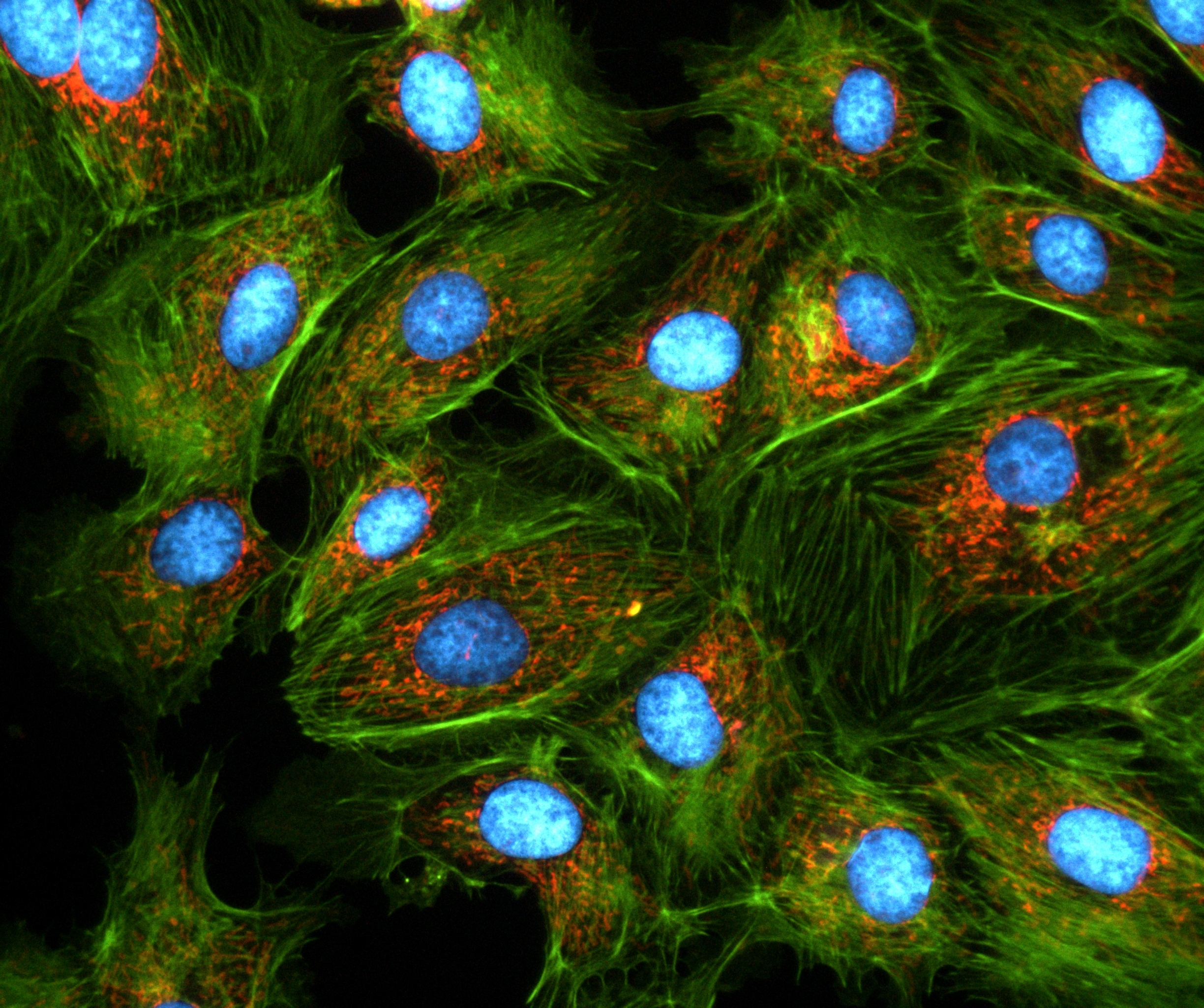
Sample : bovine pulmonary artery endothelial cell, BPAEC Filter Set : JNO-U(B), B(B), G(B) Camera : AcquCAM 23GR2 C-mount adapter : 1X Lens : UPLNAPO40X Above three images are merged and processed using ARM S/WSample : bovine pulmonary artery endothelial cell, BPAEC Filter Set : JNO-U(L), B(L), G(L) Camera : AcquCAM 23GR2 C-mount adapter : 1X Lens : UPLNAPO40X Above three images are merged and processed using ARM S/WSample : bovine pulmonary artery endothelial cell, BPAEC Filter Set : JNO-U(B), B(B), G(B) Camera : AcquCAM 23GR2 C-mount adapter : 1X Lens : PlanApo60xWLSM Above three images are merged and processed using ARM S/W
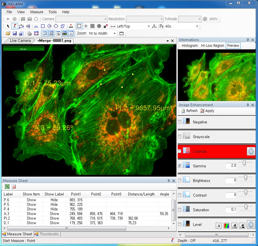
ARM is image analysis software for JNOPTIC AcquCAM cameras. This S/W is interchangeable with all WDM cameras regardless of camera brand and model and even more the most strength point is simple and easy use of length, area, angle, etc.
2. Simple measurement tools
Measurement Tools: Count, Distance, Angle, Area, etc.
Available measurement on LIVE image and saved image mode
3-1. Specialization for observation of fluorescence images
Improvement Function about Fluorescence Microscopy Image. (Used function: Auto Level, Low level, Pseudo Color, Merge Image)
Bottom on the left is merged image without improvement of image. And the bottom on the right is merged image after improvement of image. These images are shot by JNOPTIC AcquCAM Pro/G3 camera
3-2-1 Effect of high-contrast image with simple operation (Monochrome)
Improvement Function about Microscopy Image Monochrome. (Used function: Auto Level)
Effective improvement of image with simple operation. This image is shot by JNOPTIC Pro/S3 camera
3-2-2 Effect of high-contrast image with simple operation (Color)
Improvement Function about Microscopy Image. (Used function: Auto Level)
Effective improvement of image with simple operation. These images are shot by JNOPTIC AcquCAM Pro/G3 camera
3-3-1 Live Pseudo Color (for color camera)
When you get fluorescence images using color camera, if the unwanted channel was shown because of cross talk, you could remove this channel to take advantage of function of Live Pseudo.
Improvement Function about Fluorescence Microscopy Live Camera Image. (Used function: Live Pseudo Color)
The extraction of wanted color information from color image (Remove BR on RGB source) These images are shot by JNOPTIC AcquCAM Pro/G3 camera
3-3-2 Live Pseudo Color (for Monochrome camera)
When you get fluorescence images using monochrome camera, you can raise efficiency of acquisition for fluorescence images to add similar false color to the color to be shown on the eyepiece in real time.
Improvement Function about Fluorescence Microscopy Live Monochrome Camera Image. (Used function: Live Pseudo Color)
Improvement of usage environment for monochrome camera to add false color on the image. These images are shot by JNOPTIC AcquCAM Pro/S3 camera
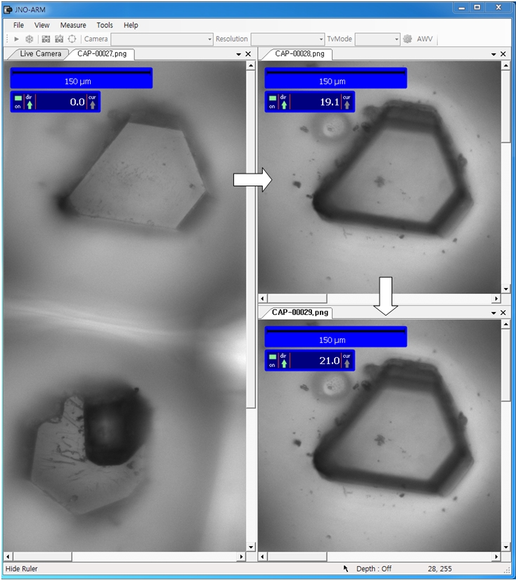
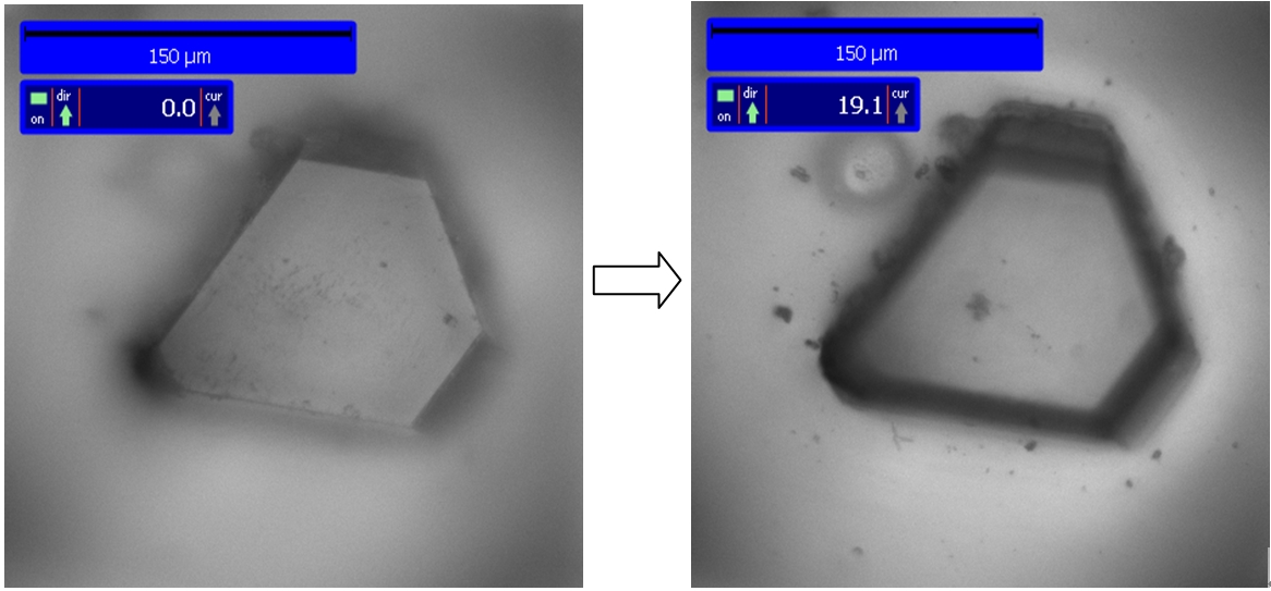
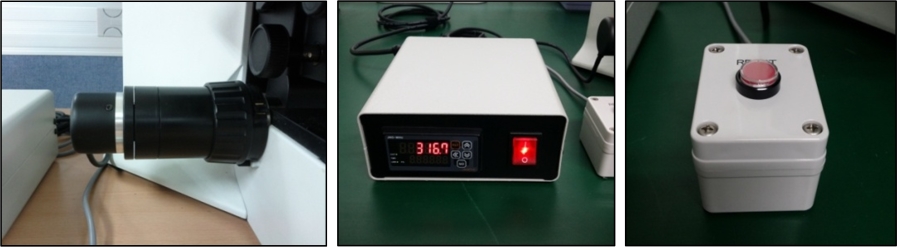
4. JNO-MHU(Option Unit)
Available measuring height with additional unit, JNO-MHU
※ JNO-MHU is sold separately as optional unit.
Left: Focus on top of sample (Z-axis reset), Right: Focus on bottom of sample (Z-axis height measurement)
< Measuring condition : over 20x objective, temp 20°C >
CA3 pyramidal neuron, IR-DIC & Alexa Fluo 488 at the same time Figure courtesy by Dr. Sooyun Kim, Seoul National University with Bx51WI, JNO-DPTS, AcquCAM 23S
JNO-DPTS CA3 pyramidal neuron, IR-DIC & Alexa Fluo 488 at the same time Figure courtesy by Dr. Sooyun Kim, Seoul National University with Bx51WI, JNO-DPTS, AcquCAM 23S
USB 3.0 Monochrome Camera
1/1.2 ” Pregius Sensor
1920×1200 pixel
Trigger input and I/O
Cost-effective mono camera for fluorescence microscope (Best)
Cost-effective camera for Department of Physiology (Best)
1/1.2 inch optimized sensor size for microscope field of view
Acquire images of optimum width combined with 0.63x adapter
AcquCAM 23S2 mono camera has a USB 3.0 interface and is the perfect solution for many industrial automation, quality assurance, security, surveillance and medical applications. The mono camera ships with the very sensitive 1/1.2 inch CMOS Pregius sensor. With up to 162 images per second, the AcuqCAM 23S2 is a low cost, yet highly versatile imaging solution. The camera includes a C to CS mount adapter, making it compatible to C and CS mount lenses.
Images with AcquCAM 23S2
Sample : bovine pulmonary artery endothelial cell, BPAEC Filter Set : JNO-U(B), B(B), G(B) Camera : AcquCAM 23S2 C-mount adapter : 1x Lens : UPlanAPO 40x Above three images are merged and processed using ARM S/W
Sample : bovine pulmonary artery endothelial cell, BPAEC Filter Set : JNO-U(B), B(B), G(B) Camera : AcquCAM 23S2 C-mount adapter : 1x Lens : UPlanAPO 40x Above three images are merged and processed using ARM S/W
Specification
Spec
Description
Size
1/1.2 inch
Resolution
H: 1920, V: 1200
Pixel size
H: 5.86 µm, V: 5.86 µm
Video formats & Frame rate
1920×1200 Y16 @ 80 fps 1920×1200 Y800 @ 162 fps
Sensitivity
0.015 lx
Exposure time
20㎲ to 30 s
Lens mount
C/Cs mount
Interface
USB 3.0
Power supply
4.5 to 5.5 VDC
Dimensions
H: 29 mm, W: 29 mm, L: 43 mm
Mass
65 g
More images with AcquCAM 23S2
Sample : bovine pulmonary artery endothelial cell, BPAEC Filter Set : JNO-U(L), B(L), G(L) Camera : AcquCAM 23S2 C-mount adapter : 1X Lens : UPLNAPO40X Above three images are merged and processed using ARM S/WSample : bovine pulmonary artery endothelial cell, BPAEC Filter Set : JNO-U(B), B(B), G(B) Camera : AcquCAM 23S2 C-mount adapter : 1X Lens : PlanApo60xWLSM Above three images are merged and processed using ARM S/W
ARM
ARM is image analysis software for JNOPTIC AcquCAM cameras. This S/W is interchangeable with all WDM cameras regardless of camera brand and model and even more the most strength point is simple and easy use of length, area, angle, etc.
Sample : bovine pulmonary artery endothelial cell, BPAEC Filter Set : JNO-U(B), B(B), G(B) Camera : AcquCAM 3G C-mount adapter : 1X Lens : PlanApo60xWLSM Above three images are merged and processed using ARM S/W
Sample : bovine pulmonary artery endothelial cell, BPAEC Filter Set : JNO-U(L), B(L), G(L) Camera : AcquCAM 3G C-mount adapter : 1X Lens : UPLNAPO40X Above three images are merged and processed using ARM S/W
Available of images with a very wide field of view
Low Noise Sensor
AcquCAM 3G color camera has a USB 3.0 interface and is the perfect solution for many industrial automation, quality assurance, security, surveillance and medical applications. The color camera ships with the very sensitive 1/1.8 inch CMOS Pregius sensor. With up to 60 images per second, the AcuqCAM 3G is a low cost, yet highly versatile imaging solution. The camera includes a C to CS mount adapter, making it compatible to C and CS mount lenses. Using the optional CS to M12 board lens adapter, the camera is also compatible to M12 board lenses.
Images with AcquCAM 3G
Sample : bovine pulmonary artery endothelial cell, BPAEC Filter Set : JNO-U(B), B(B), G(B) Camera : AcquCAM 3G C-mount adapter : 1X Lens : UPLNAPO40X Above three images are merged and processed using ARM S/W
Sample : bovine pulmonary artery endothelial cell, BPAEC Filter Set : JNO-U(B), B(B), G(B) Camera : AcquCAM 3G C-mount adapter : 1X Lens : PlanApo60xWLSM Above three images are merged and processed using ARM S/WSample : bovine pulmonary artery endothelial cell, BPAEC Filter Set : JNO-U(L), B(L), G(L) Camera : AcquCAM 3G C-mount adapter : 1X Lens : UPLNAPO40X Above three images are merged and processed using ARM S/W
ARM
ARM is image analysis software for JNOPTIC AcquCAM cameras. This S/W is interchangeable with all WDM cameras regardless of camera brand and model and even more the most strength point is simple and easy use of length, area, angle, etc.
Available of taking images with a wavelength of 650nm or more, such as CY5
High Sensitivity Sensor (Best)
Fast, seamless image transfer
Available of images with a very wide field of view
Low Noise Sensor
The displays live digital images with gradual smoothness and combines exceptional resolution with faithful color reproduction.The new sensors feature a global shutter function and able to capture a high-speed moving image without focal plane distortion. High-speed processing, low noise and low power dissipation by using column-parallel A/D conversion. equipped with trigger mode, and the external pulse can control accumulation time. The Sensor also have a pulse output function to indicate respective conditions during shutter operation and can be coordinated with peripheral circuits. High-definition images can be displayed live at a rate of high frames per second. without compression. Such imaging quality enables even the finest cellular regions to be observed clearly and distinctly without deterioration. While focusing is made stress free.
Images with AcquCAM 23GR2
Sample : bovine pulmonary artery endothelial cell, BPAEC Filter Set : JNO-U(B), B(B), G(B) Camera : AcquCAM 23GR2 C-mount adapter : 1X Lens : UPLNAPO40X Above three images are merged and processed using ARM S/W
More images with AcquCAM 23GR2
Sample : bovine pulmonary artery endothelial cell, BPAEC Camera : AcquCAM 23GR2 , Fluorescence Filter Set : JNO-U(B), B(B), G(B) Objective lens:UPLNAPO40X, C-mount adapter : 1X Above three images are merged and processed using JNO-ARM
OLYMPUS BX51, UPlanApo40x U-TV1XC
JNOpTIC AcquCAM 23GR2, JNO-UBG(B) C TYPE
OLYMPUS BX51, UPlanApo40x U-TV1XC
JNOpTIC AcquCAM 23GR2, JNO-UBG(B) B TYPE
ARM is image analysis software for JNOPTIC AcquCAM cameras. This S/W is interchangeable with all WDM cameras regardless of camera brand and model and even more the most strength point is simple and easy use of length, area, angle, etc.
2. Simple measurement tools
Measurement Tools: Count, Distance, Angle, Area, etc.
Available measurement on LIVE image and saved image mode
3-1. Specialization for observation of fluorescence images
Improvement Function about Fluorescence Microscopy Image. (Used function: Auto Level, Low level, Pseudo Color, Merge Image)
Bottom on the left is merged image without improvement of image. And the bottom on the right is merged image after improvement of image. These images are shot by JNOPTIC AcquCAM Pro/G3 camera
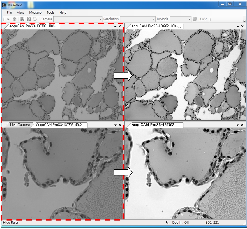
3-2-1 Effect of high-contrast image with simple operation (Monochrome)
Improvement Function about Microscopy Image Monochrome. (Used function: Auto Level)
Effective improvement of image with simple operation. This image is shot by JNOPTIC Pro/S3 camera
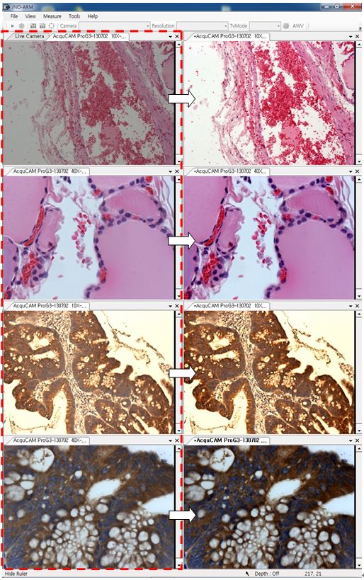
3-2-2 Effect of high-contrast image with simple operation (Color)
Improvement Function about Microscopy Image. (Used function: Auto Level)
Effective improvement of image with simple operation. These images are shot by JNOPTIC AcquCAM Pro/G3 camera
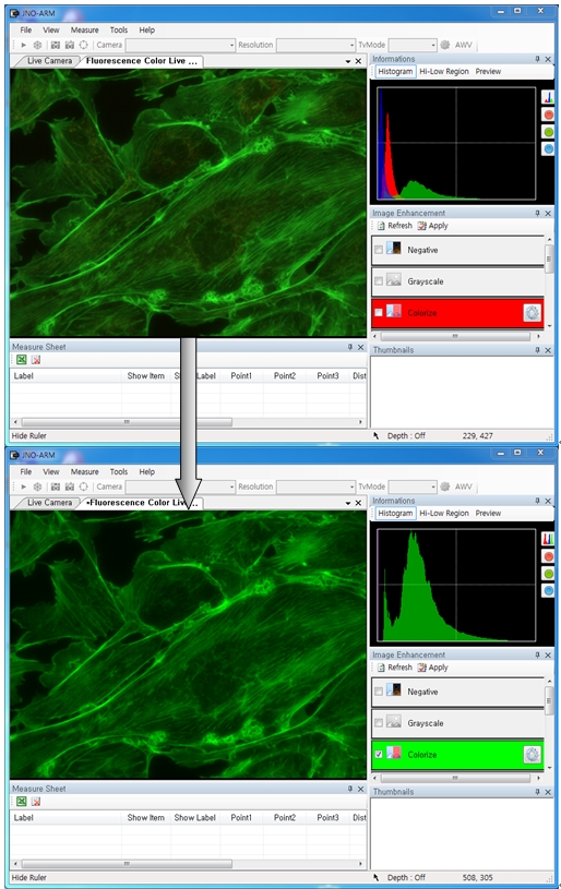
3-3-1 Live Pseudo Color (for color camera)
When you get fluorescence images using color camera, if the unwanted channel was shown because of cross talk, you could remove this channel to take advantage of function of Live Pseudo.
Improvement Function about Fluorescence Microscopy Live Camera Image. (Used function: Live Pseudo Color)
The extraction of wanted color information from color image (Remove BR on RGB source) These images are shot by JNOPTIC AcquCAM Pro/G3 camera
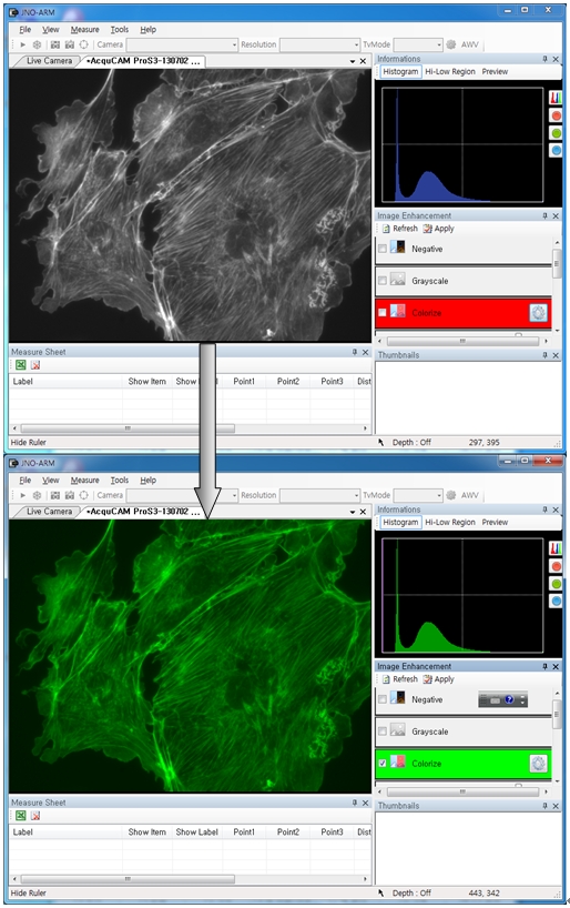
3-3-2 Live Pseudo Color (for Monochrome camera)
When you get fluorescence images using monochrome camera, you can raise efficiency of acquisition for fluorescence images to add similar false color to the color to be shown on the eyepiece in real time.
Improvement Function about Fluorescence Microscopy Live Monochrome Camera Image. (Used function: Live Pseudo Color)
Improvement of usage environment for monochrome camera to add false color on the image. These images are shot by JNOPTIC AcquCAM Pro/S3 camera
4. JNO-MHU(Option Unit)
Available measuring height with additional unit, JNO-MHU
※ JNO-MHU is sold separately as optional unit.
Left: Focus on top of sample (Z-axis reset), Right: Focus on bottom of sample (Z-axis height measurement)
< Measuring condition : over 20x objective, temp 20°C >
Available of images with a very wide field of view
Low Noise Sensor
AcquCAM 5G color camera has a USB 3.0 interface and is the perfect solution for many industrial automation, quality assurance, security, surveillance and medical applications. The color camera ships with the very sensitive 2/3 inch CMOS Pregius sensor. With up to 38 images per second, the AcuqCAM 5G is a low cost, yet highly versatile imaging solution. The camera includes a C to CS mount adapter, making it compatible to C and CS mount lenses. Using the optional CS to M12 board lens adapter, the camera is also compatible to M12 board lenses.
Images with AcquCAM 5G
Sample : bovine pulmonary artery endothelial cell, BPAEC Filter Set : JNO-U(B), B(B), G(B) Camera : AcquCAM 5G C-mount adapter : 1X Lens : UPLNAPO40X Above three images are merged and processed using ARM S/WSample : bovine pulmonary artery endothelial cell, BPAEC Filter Set : JNO-U(B), B(B), G(B) Camera : AcquCAM 5G C-mount adapter : 1X Lens : PlanApo60xWLSM Above three images are merged and processed using ARM S/WSample : bovine pulmonary artery endothelial cell, BPAEC Filter Set : JNO-U(L), B(L), G(L) Camera : AcquCAM 5G C-mount adapter : 1X Lens : UPLNAPO40X Above three images are merged and processed using ARM S/W
ARM is image analysis software for JNOPTIC AcquCAM cameras. This S/W is interchangeable with all WDM cameras regardless of camera brand and model and even more the most strength point is simple and easy use of length, area, angle, etc.


