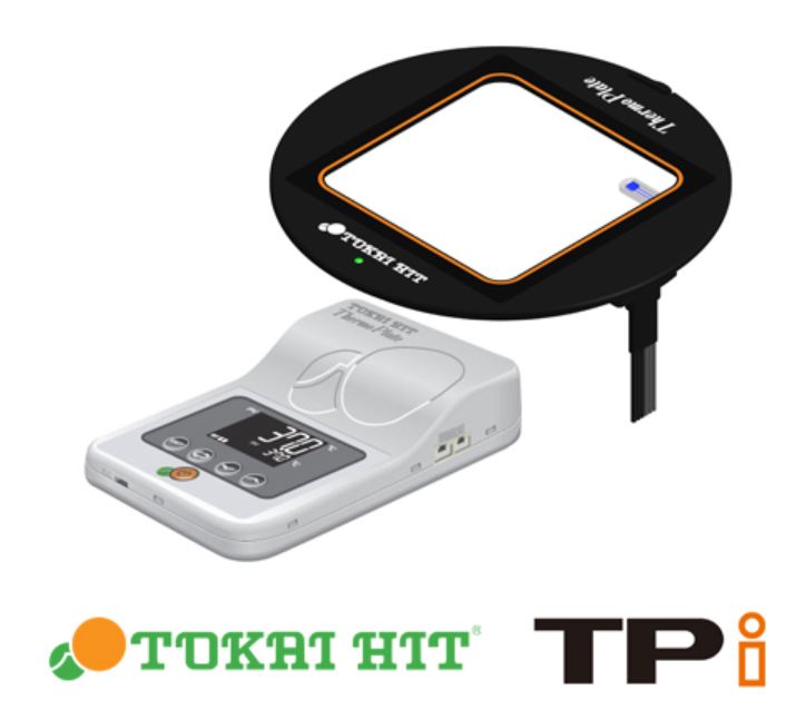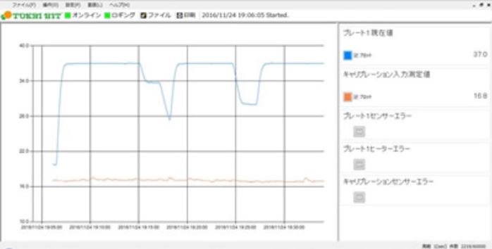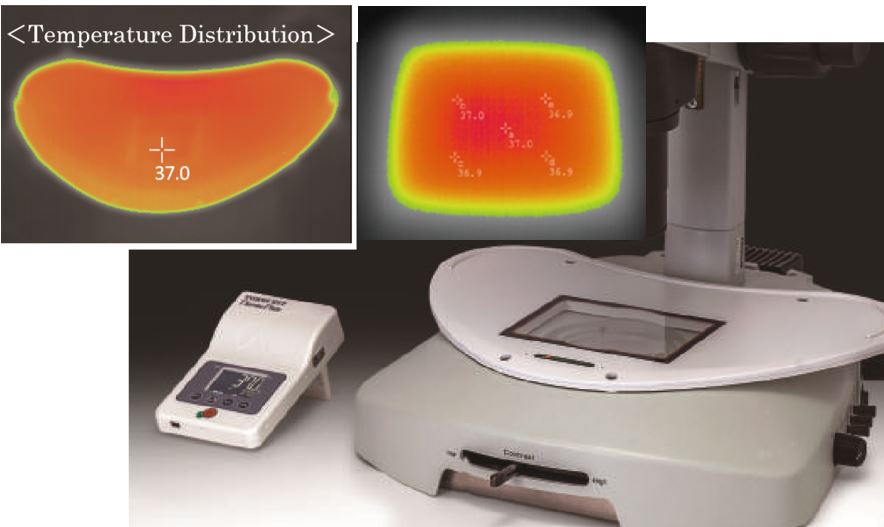
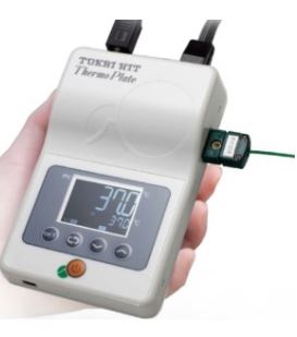
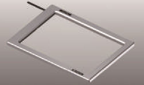
TOKAI HIT Thermo Plate
Clear Glass Heater For Olympus
Nikon, Zeiss, Leica, Keyence 등 각종 메이커 가능합니다. 문의 바랍니다.
온도 범위 : 상온 ~ 60 ℃ (TPi-UNIX 제외)
“X”로 끝나는 모델은 강화 유리 적용 모델 입니다.
(TPi-UNIX → 강화유리 적용 모델)
공통 기본 구성품
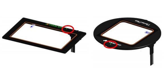
- Heating Plate
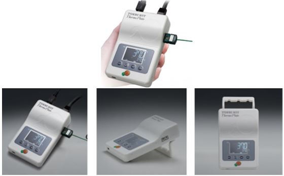
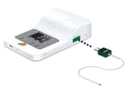
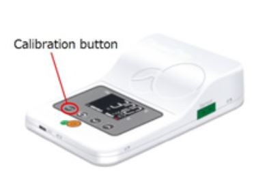
- TPi Controller
- Mounting Hook
- Power code
- AC Adapter
- Temperature Sensor (TPiE 모델 제외)
- Extension Wire (TPiE 모델 제외)
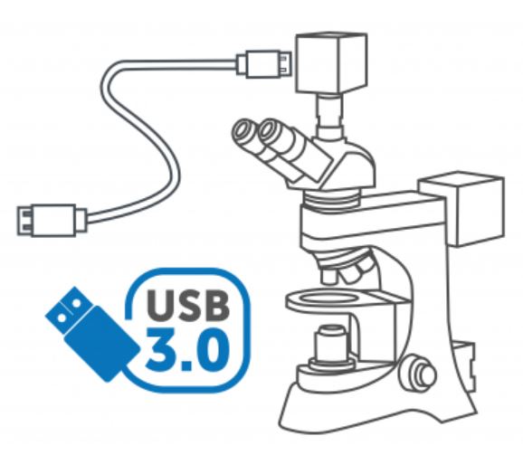
- TEM Install CD&USB cable (TPiE 모델 제외)
1. TPi-110RX


Microscope: Olympus IX83 / IX 73 / IX81 / IX71 /IX 51 / IX70/ IX50, IMT2
Applicable stage: Cross stage with 110 mm round opening
- Setting range: ambient ~ 60℃
- Plate dimension: φ110 mm
- Heating area: W70 × D70 mm
- Glass thickness: 0.5 mm
2. TPi-110R13


Microscope: Olympus IX83 / IX 73 / IX81 / IX71 /IX 51 / IX70/ IX50, IMT2
Applicable stage: Cross stage with 110 mm round opening
- Setting range: ambient ~ 60℃
- Plate dimension: φ110 mm
- Heating area: W70 × D70 mm
- Glass thickness: 1.3 mm
- Ideal for relief contrast observation with a glass bottom dish
3. TPi-IX3X


Microscope: Olympus IX83 / IX 73
Applicable stage: XY manual (IX3-SVR)/XY motorized (IX3-SSU) stage
- Setting range: ambient ~ 60℃
- Plate dimension: W189.5 x D155.5 mm
- Heating area: W174 × D127 mm
- Glass thickness: 0.5 mm
4. TPi-IX3-13

Microscope: Olympus IX83 / IX 73
Applicable stage: XY manual (IX3-SVR)/XY motorized (IX3-SSU) stage
- Setting range: ambient ~ 60℃
- Plate dimension: W189.5 x D155.5 mm
- Heating area: W155 × D130 mm
- Glass thickness: 1.3 mm
5. TPi-SQX


Microscope: Olympus IX series
Applicable stage: manual/motorized XY stage with 160 × 110 mm aperture for Prior Scientific stage H117
아래 아답타를 사용하여 다양한 스테이지에 설치 가능
- MK-IX3 (OLYMPUS IX3-SSU, IX3-SVR)
- MK-SIG (OLYMPUS BX3-SSU, IX2-SFR, IX2-SVL2)
- TI2-ZILCS (NIKON TI2-S-SE-E, TI2-S-SS-E)
- TI2-RA (NIKON TC-S-SR, TC-S-SRF)
- TID-ZILCS (NIKON TI-S-E, TI-S-ER)
- MK-RA (NIKON TI-SR, TI-SSR)
- MK-EVOS (Thermo Fisher Scientific EVOS FL)
- MK-EVOS-AT (Thermo Fisher Scientific EVOS FL Auto)
- Setting range: ambient ~ 60℃
- Plate dimension: W160 x D110 mm
- Heating area: W128 × D84 mm
- Glass thickness: 0.5 mm
6. TPi-CKX53X


Microscope: Olympus CKX53 series
Applicable stage: XY mechanical stage CKX3-MVR
- Setting range: ambient ~ 60℃
- Plate dimension: W190 x D138 mm
- Heating area: W174 × D127 mm
- Glass thickness: 0.5 mm
7. TPi-CKX


Microscope: Olympus CKX41 / CKX31 / CK40 / CK30 / CK2
Applicable stage: Olympus XY mechanical stage IX-MVR
- Setting range: ambient ~ 60℃
- Plate dimension: W127 x D85 mm
- Heating area: W103 × D63 mm
- Glass thickness: 0.5 mm
8. TPi-CKTS


Microscope: Olympus CKX41 / CKX31, CK40 / CK30 / CK2 Nikon TS / TS-100
Applicable stage: Olympus XY mechanical stage IX-MVR, Nikon XY mechanical stage TE/TI-SAM
- Setting range: ambient ~ 60℃
- Plate dimension: W150 x D117 mm
- Heating area: W131 × D95 mm
- Glass thickness: 0.5 mm
9. TPi-SX


Microscope: Upright Microscopes BX53 / BX43 / CX40 / CH40 / CH30
Applicable stage: Mechanical stage for upright microscopes
- Setting range: ambient ~ 60℃
- Plate dimension: W142 x D115 mm
- Heating area: W128 × D95 mm
- Glass thickness: 0.5 mm
10. TPi-SZX2X


Microscope: Olympus SZX16 / SZX10
Applicable illumination base: SZX2-ILLB / SZX2–ILLD / SZX2- ILLK / SZX2- ILLT / SZX2- ILLTQ / SZX2- ILLTS
- Setting range: ambient ~ 60℃
- Plate dimension: W238 x D227 mm
- Heating area: W162 × D152 mm
- Glass thickness: 1.0 mm
11. TPi-SZX1


Microscope: Olympus MVX10 SZX12 / SZX9 / SZX7
Applicable illumination stand: SZX-ILLK / SZX-ILLB2 / SZX-ILLD2
- Setting range: ambient ~ 60℃
- Plate dimension: W205 x D205 mm
- Heating area: W170 × D170 mm
- Glass thickness: 1.0 mm
12. TPi-SZ2


Microscope: Olympus SZX7, SZ61
Applicable illumination base: SZ2-ST + SZ2-ILA
- Setting range: ambient ~ 60℃
- Plate dimension: W278 x D175 mm
- Heating area: W230 × D146 mm
- Glass thickness: 1.0 mm
13. TPi-OZX


Microscope: Olympus SZ60 / SZ40 / SZ11
Applicable illumination stand: OLYMPUS SZ60 / SZ40 / SZ11 illumination stand
- Setting range: ambient ~ 60℃
- Plate dimension: W180 x D230 mm
- Heating area: W162 × D152 mm
- Glass thickness: 1.0 mm
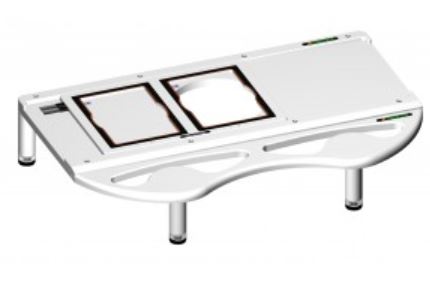
14. TPi-UNIX


Universal Type for Stereo Microscope
For Various types of illumination base
- Setting range: ambient ~ 50℃
- Plate dimension: W435 x D220 mm
- Legs adjustable range : 75~100mm
- Heating area: W400 × D175 mm
- Glass thickness: 1.5 mm
15. TPi-W / TPi-WL
Universal Large Type (W230 x D180 / W310 x D220)
- Setting range: ambient ~ 60℃
- Plate dimension: W230 x D180 / W310 x D220 mm
- Heating area: W180 × D140 / W250 x D170 mm
- Glass thickness: 1.5 mm
16. TPiD-VITX
Universal Type for stereo microscopes
For Thawing Process of Frozen embryo
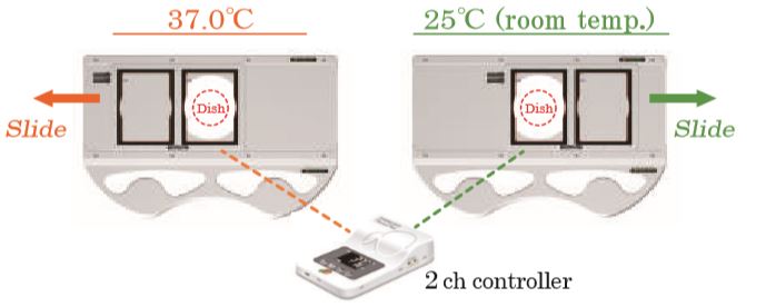
- Setting range: ambient ~ 60℃
- Base dimentions:W435 × D280 mm
- Plate dimentions: W230 × D148 mm
- Legs adjustable range:75 – 100 mm
- Heating area:W95 × D128 × 2 pcs
- Glass thickness:0.5 mm
Thermo Plate / Thermo Stage / Warming Plate / Heating Plate / Heating Stage / 가열 스테이지