J.H.JIN의 모든 글

F-view II

F-view II

F-View II 는 12-bit monochrome CCD 카메라 입니다.

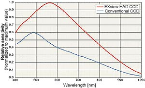
이 제품은 극도로 높은 감도, 저 노이즈, 고해상도의 정량화할 수 있는 이미지를 요구하는 응용분야에 이상적입니다.

F-View IIFireWire 기술 (IEEE1394)에 의한 Peltier 냉각 및 전력을 공급받으며 FISH 혹은 Single band-pass 필터를 사용하는 다중 노출 형광 응용분야를 위해 특별히 설계되었습니다.

F-View II 는  “analySIS Five” Image Analyzer Software에 완전히 통합됩니다.

Digital solutions – Developed to be exceptional!

Soft Imaging System’s digital cameras are designed to meet the steadily rising demands on digital image acquisition for all areas of microscopy. And so is the newly developed 12-bit, cooled CCD color camera F-View II, our latest 12-bit, Peltier-cooled, FireWire™-driven solution for fluorescence microscopy.

The search mode enables you to rapidly locate and conveniently focus in on the area of your sample of interest. This prevents any unnecessary bleaching of the sample.

The camera will automatically switch over to the high-resolution mode for acquisition. F-View II can also be easily operated using a laptop via the FireWire™ port.

All camera functions are fully controlled by the analySIS® image-processing software. Real-time functions enable the use of the entire dynamic range under all conditions and guarantee the best contrast.

F-View II’s integration into analySIS® provides all the capabilities and advantages of state-of-the-art digital image processing and analysis, ranging from image labeling, archiving, report generation and e-mailing, as well as photo-realistic printouts without the darkroom.

 High Resolution
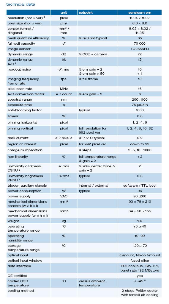
The CCD chip of the F-View II has 1376 x 1032 pixel resolution. The 12-bit dynamic range means that even acquisition of images with very bright and faint regions is dynamically optimal. 


 High sensitivity
CCD elements are so sensitive that they have no trouble detecting even the weakest of signals. The electronic shutter offers variable exposure times ranging from 100 µs to 160 seconds.


 High frame rate
The high speed ADC (Analog-Digital Converter) working at a clock rate of 20 MHz in full 12-bit dynamic range is able to perform double sampling even at a readout rate of 20 MHz. Various frame rates are supported by this camera. For example, the camera can be set to acquire at a high frame rate of more than 22 fps at TV resolution using 2x binning. View your zoomed-in sample, locate the area of interest and focus – all conveniently onscreen. No longer are you forced to trade off speed for quality. For acquisition the system switches automatically into the high-resolution mode. This avoids bleaching of your fluorescence specimen and offers optimal performance when setting parameters.


 FireWire™ Interface (IEEE 1394)
FireWire™ technology guarantees that the F-View II installation is easy on any PC or laptop equipped with a FireWire™ port. The days when you were limited to a frame grabber and just a single camera are history. FireWire™ technology enables you to use multiple cameras on the same PC.


 Peltier-Cooling
Image noise is generally the result of the CCD chip not being cooled to a low enough temperature (“dark current”). For the F-View II, thermal noise is not a problem because the CCD chip is Peltier-cooled and stabilized at 10°C. Noise is further suppressed by the application of a highly efficient digital readout technique known as Correlated Double Sampling (CDS).


 Compact design
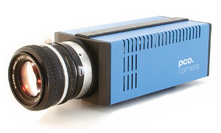
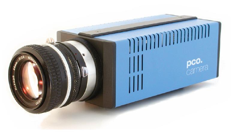
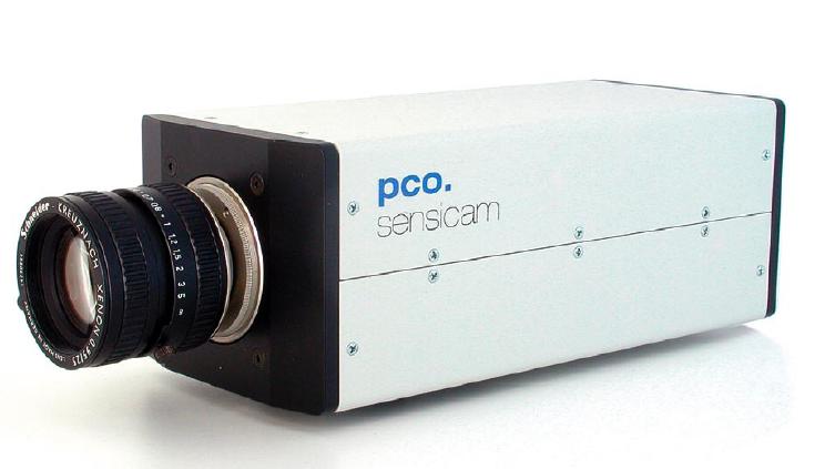
With its elegantly compact, newly designed housing, the F-View II can be mounted onto all light microscopes with a C-mount adaptor. No other interfaces or adaptors are necessary. Plus, you only need one cable for getting data and power to the PC’s FireWire™ port. No more clutter and no “octopus” of cables getting in your way.

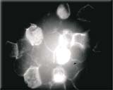
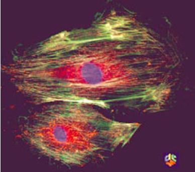
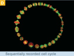
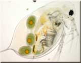
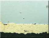
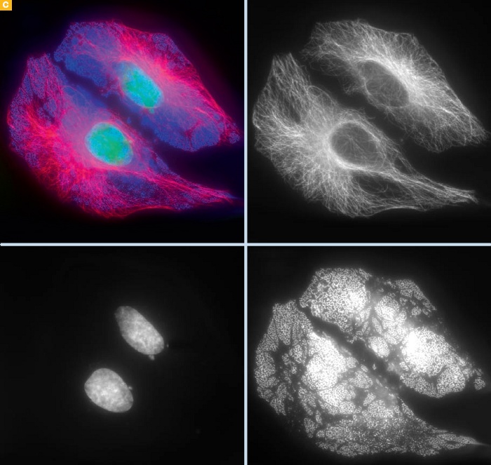
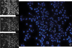
DAPI, PE, APC, PerCP & FITC image acquisitions
of stem cells: combined acquisitions done via themFip analySIS® add-in

source: Dr. Hans Wessels
University Clinic for Children, Tübingen, Germany

 Real-time functions
F-View II is fast. Put this in combination with the high speed of today’s CPU’s, and you’ve got an attractively broad range of real-time functions within analySIS®available to you. These include automatic contrast control, sharpness monitoring and histogram display.


 Analyzing images
A vast library of text, graphic and editing functions is available for labeling images. Special filters and professional particle analysis assist in more extensive investigation of images. All this makes it simple to obtain reliable and reproducible results quickly.


 No more darkroom
Photo-quality printouts can be obtained following acquisition without any need for a darkroom nor its developing chemicals. And these photo-quality printouts are in your hands in minutes.
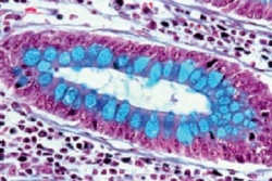
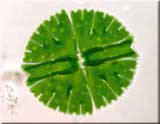
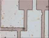
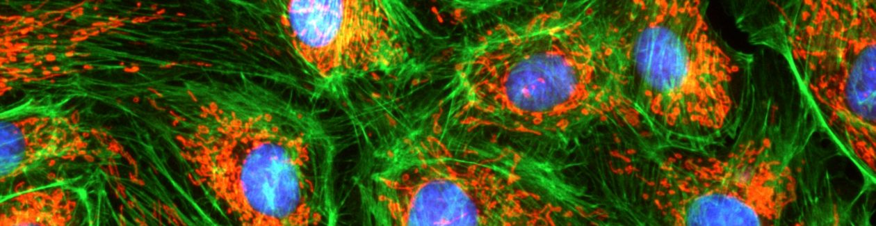
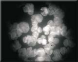
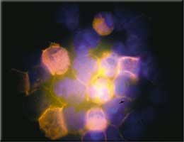
a) Her2 gene (Spectrum Orange)
b) DNA (DAPI)
c) Chromosome (Spectrum Green)
d) Combination with
mFip
Specimen: Norbert Wey
University of Zürich
Department of Pathology
Switzerland

Archiving
analySIS® offers you a powerful, fully networkable, image-archiving system that handles all the images and data generated during the process of image acquisition and documentation. Images can be stored along with text, sheets and other graphics as records for complete documentation of tasks and processing steps. Input, display and query masks can be defined independently.

Automatic report generation
Now you can produce multi-page reports quickly and efficiently. Select multiple images in the image database and insert them all into the report via a single command. In addition to the images themselves, you can have information from any database field automatically included in reports. Automatic scaling, detail zooms, and more – all available to optimize the way you work with images.

All documents generated using the analySIS® software can be inserted into reports. Use the report generator to print out images, related measurement sheets, and diagrams – all on the same page. This report generator provides you with the utmost flexibility for page layout and design. You set up your own templates exactly the way you want them to be. Templates need to be created just once. Templates are what your reports are based on and ensure that the appearance of your documents is uniform. Use the RTF Export function to export your reports to MS Word for continued editing.

Software control of all camera functions

   Manufacturer programmable RISC CPU + FPGA via firmware download
   Multi-threading code support on multi-CPU PC


Depending on options, analySIS® expansion level and

   Real-time automatic contrast control

   Real-time automatic white balance

 Real-time shading correction

 Real-time sharpness monitor

   Real-time histogram display

   Real-time FFT computation and display
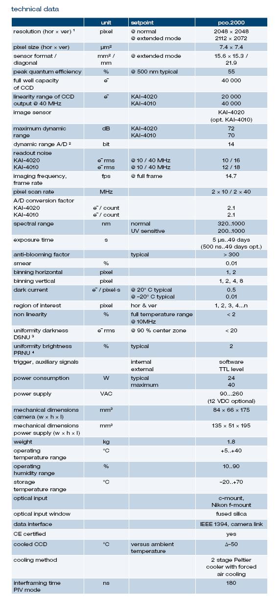
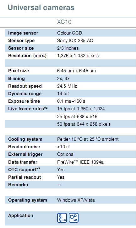
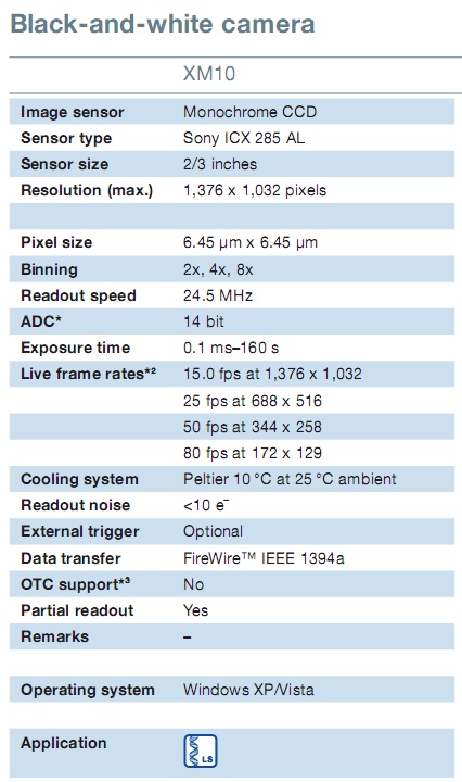
 Specifications

Image Device 2/3 inch monochrome CCD Sensor
(Effective area 8.9 x 6.7 mm array)
Effective Pixels 1376 x 1032 pixel, 6.45 um square pixels
Frame Rate > 22 fps @ 2x binning
39 fps @ 4x binning
72 fps @ 8x binning
Binning 2x, 4x, 8x
Dynamic Range 12 bit
Exposure 100 us ~ 160 sec
Cooling Peltier cooled, 10℃ @ 25℃ ambient
Pixel clock rate 20 MHz
Readout noise < 2 counts
Non-linearity < 0.6%
Anti-blooming > 300
Dimensions (W x H x D) 100 x 85 x 50 mm
Mass 570g
Temperature monitor CCD chip & housing
Temperature stabilized Yes, ± 0.5 ℃
Interface Connector FireWire (IEEE1394)
Lens mount Standard C-mount

Pixelink PL-B686

Pixelink PL-B686 카메라는 고성능 현미경을 위하여 고안되었으며 고 품질, 고 해상력 영상을 획득하도록 디자인 되었습니다.

가장 큰 특징인 6백6십만 화소1인치형 CMOS 센서는 최고 해상도에서 5프레임의 속도 혹은 88프레임의 VGA 해상도의 영상 보기를 제공합니다.

다른 특징으로는 일부 영역의 영상 발췌 및 2x, 3x, 4x 및 6x Binnnig 기능을 제공합니다.

PL 686 카메라는 i-solution capture 소프트웨어 패키지와 함께 명시야 현미경 영상을 위한 높은 생산성을 갖는 영상 분석 시스템이 됩니다.

i-solution captures는 측정 및 주석, 영상의 개선과 획득한 영상의 관리 및 DB 저장과 같은 카메라를 위한 핵심 기능들을 제공합니다.

Specification Sheet

Pixelink PL-B686C
  Color/ MonoColor
  Resolution3000×2208
  Frame Rate at Full Resolution5
  Sensor Type1″ CMOS
  Lens formatC-mount
  Pixel size3.5 um x 3.5 um
  Sensor Diagonal13.1 mm
  Bit Depth8 or 10
  Shutter TypeRolling
  Dynamic Range56 dB
  InterfaceUSB2.0

AcquCAM Pro

TOMORO AcquCAM Pro
– Discontinued

TOMORO ACQUCAM PRO는 전문적인 현미경용

디지털 카메라로 완벽한 해상력의 이미지를 제공

합니다.

CMOS보다 더 정교한 이미지를 제공하는 CCD이며

130만 화소의 고화질 이미지를 제공합니다. 

또한 IEEE 1394 규격을 지원으로 사용상의 편리함과

400Mbps의 빠른 전송 속도로 고화질의 이미지를

초당 15프레임의 속도로 관찰 할 수 있습니다.

Features

* 1.3 Mega pixel Sony ICX205AK CCD Sensor

* Color, Progressive scan

* Lower Fixed Pattern Noise

* High Speed IEEE1394(400Mbits/sec)

* 15 fps at 1280×960 resolution

* Auto White Balance support

* RGB Bayer video output

(?)Select 8or 10-bit pixel data

(?)Faster frame rates with reduced Region of Interest

* GPI/Os for control of peripherals and synchronization of lighting (4in/4out)

* Preview Software provided (TOMORO Capture)

* Software compatible with Windows 98SE/2K/XP operating systems

* TWAIN and Direct Show Compatible

Specifications

SensorSony ICX205AK
TypeCCD interline transfer, progressive scan
Size1/2 inch
ResolutionH:1280, V960
Pixel sizeH:4.65um, V:4.65um
Video formats &(Frame rates)Upto 15 Fps
UYUV 1280 x 960(3.75 / 7.5images/sec)
Y800 1280 x 960(3.75 / 7.5 / 15images/sec)
Sensitivity19 IX at 1/30s, gain Ø dB
Exposure time2 to 1/20000sec
Gain0 to 36 dB
White balanceAuto / Manual
Lens mountC/Cs mount
InterfaceIEEE 1394, 2 ports, 6 pins 400Mbit/s
Power supply8 to 30 VDC via IEEE
DimensionsH:50mm, W:50mm, L:55mm
MassApprox 300g

AcquCAM II

TOMORO AcquCAM II
– discontinued

TOMORO ACQUCAM II 는 뛰어난 가격대

성능비를 지닌 현미경용 이미지 분석에 적합한

DIGITAL CAMERA입니다.

130만 화소가 제공하는 고화질의 이미지는

전용 On-board 프로세서에 의해 더욱 정교한

이미지를 제공합니다.

또한 USB2.0 규격을 지원으로 사용상의 편리함과

480Mbps의 빠른 전송 속도로 고화질의 이미지를

초당 15프레임의 속도로 관찰 할 수 있습니다.

Features

* 1.3 Mega pixel Image Sensor

* Color, Progressive scan

* Lower Fixed Pattern Noise

* High Speed USB2.0 (4.8Mb/s)

* 15 fps at 1280×1024 resolution, 60 fps at 640×480 resolution

* Auto White Balance support

* Snap-shot mode for use with strobe

* RGB Bayer video output

* Select 8or 10-bit pixel data

* Faster frame rates with reduced Region of Interest

* GPI/Os for control of peripherals and synchronization of lighting (4in/4out)

* Preview Software provided (TOMORO AcquPro 2005)

* Software compatible with Windows 98SE/2K/XP operating systems

* TWAIN and Direct Show Compatible

Specifications

Image Device1/2 inch Color CMOS Sensor (6.5 x 5.3 mm array)
Effective Pixels1280×1024, 5.2 um square pixels
Frame Rate15 fps at 1280Í1024, 60fps at 640×480
SensitivityHigh
Dynamic Range60dB
Auto ExposureAutomatic / Manual
White BalanceAutomatic / Manual
Dimensions (W x H x D)7.5 x 7.5 x 6.5 cm
MassApprox 300g
Power RequirementUSB bus power, or external 6V DC, 500mA
Power Consumption~ 2.5 Watts
Operating Temperature0 ℃ ~ +50 ℃
Operating Humidity20 % ~ 80 % Non-Condensing
Interface ConnectorStandard USB (USB 2.0)
Lens mountC-mount
Frame BufferInternal 3 Frame buffer (Optional expansion to 12 frames)

PCO. 2000

PCO. 2000

cooled digital 14bit CCD camera system


PCO사에서 제작한 PCO.2000 카메라는 14bit cooled CCD 시스템으로 400만화소(20482048)의 고해상도로 이미지를 보여줍니다.

 camRAM 이라는 새로운 이미지 memory를 사용하여 160 MB/s 의 빠른 이미지 레코딩을 구현합니다.

 compact 카메라와 external power supply 로 구성되어 있으며, 이미지 데이터는 컴퓨터로 standard data interface(IEEE 1394 firewire, camera link)를 통해 전송됩니다.

PCO.2000의 디지털 카메라 시스템은 저광량과 piv camera application 분야에 완벽하게 적합합니다.

Features

  ▲ excellent resolution (2048 × 2048 pixel)

  ▲ 14 bit dynamic range

  ▲ frame rate of 14.7 fps at full resolution

  ▲ image memory in camera (camRAM up to 4 GB)

  ▲ excellent low noise of 10 e¯ rms @ 10 MHz

  ▲ thermo-electrical cooling of -50° C vs. ambient

  ▲ standard interfaces (IEEE 1394, camera link)

  ▲ UV sensitive & color CCD image sensor available


Quantum Efficiency


Specification

PCO. 4000

PCO. 4000

cooled digital 14bit CCD camera system


PCO사에서 제작한 PCO.4000 카메라는 14bit cooled CCD 시스템으로 천만화소(40082672)의 고해상도로 이미지를 보여줍니다.

 camRAM 이라는 새로운 이미지 memory를 사용하여 128 MB/s 의 빠른 이미지 레코딩을 구현합니다.

 compact 카메라와 external power supply 로 구성되어 있으며, 이미지 데이터는 컴퓨터로 standard data interface(IEEE 1394 firewire, camera link)를 통해 전송됩니다.

PCO.4000의 디지털 카메라 시스템은 microscopy, aerial photography나 quality control과 같이 저광량과 고해상도의 application 분야에 최고로 적합합니다.

Features

  ▲ superior resolution (4008 × 2672 pixel)

  ▲ 14 bit dynamic range

  ▲ frame rate of 5 fps at full resolution

  ▲ image memory in camera (camRAM up to 4 GB)

  ▲ excellent low noise of 12e¯ rms @ 8 MHz

  ▲ thermo-electrical cooling of -45° C vs. ambient

  ▲ standard interfaces (IEEE 1394, camera link)

  ▲ UV sensitive & color CCD image sensor available


Quantum Efficiency


Specification


Examples of applications


Sensicam QE

Sensicam QE

cooled digital 12bit CCD camera system

PCO사에서 제작한 Sensicam QE 카메라는 고성능 cooled digital 12bit CCD 로 보다 향상된 electronic technology 시스템입니다.

  열전자 cooling 센서는 이미지의 noise를 매우 적게 줄여주며 고감도로 작동하게 해줍니다.

  뛰어난 Quantum efficiency (up to 62%)를 자랑하며 고속의 serial data link(fiber optic link available)로 PC에 연결됩니다.

Sensicam QE 저광량 카메라 시스템은 Fluorescence imaging, confocal & light microscopy 와 같이 고감도이며 저잡음을 필요로하는 분야에 최고로 적합합니다.

Features

  ▲ super quantum efficiency up to 65%

  ▲ extremely low noise, down to 4e- rms

  ▲ 12bit dynamic range

  ▲ thermo-electrical cooling (Peltier) down to -12°C

  ▲ high resolution (1376 x 1040pixel)

  ▲ shutter / exposure times from 500ns – 1000s

  ▲ binning (horizontal & vertical)

  ▲ region of interest (ROI)

  ▲ 10 frames per second at full CCD resolution

  ▲ remote control up to 1500m distance (with fiber optic link)

  ▲ free software camware included


Quantum Efficiency


Specification


Examples of applications

Fluorescence in-situ hybridization (FISH) of an endothel cell.


Sensicam EM

Sensicam EM

electron multiplication digital 12bit CCD camera system

PCO사에서 제작한 Sensicam EM 카메라는 Electron Multiplication  CCD 센서를 사용하여 보다 고감도의 영상을 제공합니다.

  12 bit의 cooled digital CCD는 상당히 적은 광량의 어플리케이션에서 강력한 성능을 발휘합니다.

  뛰어난 Quantum efficiency (up to 65%)를 자랑하며 고속의 serial data link(fiber optic link available)로 PC에 연결됩니다.

이 저광량 카메라 시스템은 scientific image(e.g. microscopy, biomarker and label imaging) 과 night vision 의 분야에 활용이 매우 적합합니다.


Features

  ▲ Electron multiplication gain of up to 1000

  ▲ superior resolution (1004 × 1002 pixel) for EMCCD

  ▲ extremely low noise < 1e ¯

  ▲ excellent quantum efficiency of up to 65 %

  ▲ 12 bit dynamic range

  ▲ shutter / exposure times from 75 μs to 1 h

  ▲ thermo-electrical cooling of -45°C vs. ambient

  ▲ free software camware and software development kit included


Quantum Efficiency


Specification


XC10

OLYMPUS – XC10

뛰어난 이미지 품질, 외부 트리거 기능과 장 노출 시간 – 이것들은 이 펠티어 냉각 칼라 카메라가 제공하는 많은 속성중의 단지 일부 입니다.

XC101,376 x 1,032  픽셀의 CCD 칩을 내장하고 각 칼라 채널은 14 비트를 제공합니다. 2x 비닝 모드로 카메라는 초당 15 프레임보다 고속의 영상을 제공합니다.

이것은 XC10을 고속 영상 획득을 요구하는 어플리케이션에 대한 이상적인 카메라임 말합니다.  게다가 빠른 영상 전환은 샘플의 포커싱이나 관심 영역을 PC 스크린 상에서 찾을 수 있게 합니다.

이러한 CCD 센서에 의한 우수한 특성들은 펠티어 냉각과 결합하여 매우 낮은 잡음의 풍부한 디테일과 컨트라스트를 갖는 칼라 이미지를 가능하게 하며 최대 160초의 매우 긴 노출 시간을 설정 가능하게 합니다. 이는 카메라가 일반적인 형광 어플리케이션의 약한 신호 강도를 감지하는데 사용될 수 있습니다.

고 감도 (High sensitivity)

XC101,376 x 1,032 픽셀의 해상도를 가집니다. 고 효율의 판독 기술 (더블 샘플링)은 CCD 센서의 펠티어 냉각과 결합하여 우수한 신호 대 잡음비의 이미지를 생성합니다. 고 감도 CCD 소자는 희미한 신호조차도 감지할 수 잇습니다. 전자 셔터는 0.1 milisecond 에서 160초에 이르는 범위의 다양한 노출 시간을 제공합니다.

고속 (High Frame rate)

전 14 비트의 동적 범위의 24MHz의 클럭 속도에서 작동하는 고속 ADC (아날로그 디지털 변환기)는 24.5MHz의 판독 속도에서도 더블 샘플링을 수행할 수 있습니다. 댜앙한 프레임 속도가 지원됩니다. 예를 들어, 2x 비닝 기능을 사용하여 TV 해상도에서 15 프레임 보다 고속의 획득을 설정할 수 있습니다. 편리하게 스크린상에서 포커스와 원하는 영역의 위치를 확대된 샘플을 볼 수 있습니다. 더 이상 품질을 위하여 속도를 떨어뜨릴 필요가 없습니다. 이는 형광 시편의 Bleaching을 막고 최적의 설정을 제공합니다.

daphnia longispina
(long-spined water flea) with eggs
micrasterias rotata
(a desmid – single-celled green algae)
stem axis of tulipa gesneriana
(garden tulip)
carbon fiber
chip structure
protective lacquer coating

  Life Science 에 적합함을 나타냅니다.
  Material Science 에 적합함을 나타냅니다.


XC10 Specification

XM10

OLYMPUS – XM10

XM10는 신뢰할수 있는 형광 현미경 이미지를 제공하는데 필요한 고 분해능,  고감도, 냉각 CCD 소자, 가변 해상도 및 옵션의 외부 트리거 기능들을 제공합니다.

올림푸스 XM10은 훌륭한 기능과 표준 C-mount를 이용함으로 어떠한 현미경에도 쉽게 장착할 수 있습니다.

XM10은 OLYMPUS ‘Cell*’ 소프트웨어 및 ‘analySIS’ 소프트웨어를 완벽히 지원 합니다.

험을 위한 올바른 선택

XM101,376 x 1,032 픽셀의 14비트 분해능의 냉각 CCD 소자를 사용합니다. ( 25 ˚C 상온에서 10 ˚C 로 냉각)

최대 해상도에서 15 프레임의 영상 전송이 가능하며 라이브 모드에서의 감도를 향상 시키고 뛰어난 프레임 속도를 위한 2x, 4x, 8x의 3가지 binning 모드를 지원합니다.

이는 고 감도 형광 샘플의 형광을 유지하는 동안 초점을 쉽게 맞추고 뷰 영역에서의 관심 영역을 쉽게 찾도록 합니다.

  • 25 fps at 688 x 516 pixels (2x binning)
  • 50 fps at 344 x 258 pixels (4x binning)
  • 80 fps at 172 x 129 pixels (8x binning)

  4가지 모델

XM10은 핵심 기능을 반영하기 위하여 4가지 다른 모델을 선택 가능합니다.

기본 XM10을 확장한 Ir 확장 모델 “XM10-Ir“은 CY5와 CY7과 같은 near-Ir (근 적외선)을 포함한 형광 염료의 전 영역에 대하여 완벽하게 동작합니다.

외부 하드웨어 트리거 “XM10-T” 모델은 Cell^M 이나 Cell^R을 통하여 사용자가 원하고자 할 때의 정확한 이미지 획득을 가능하게 합니다.

이 두 가지 속성을 합친 “XM10-TIr”은 형광 현미경상의 모든 표준들을 위한 완벽한 카메라가 될 것입니다.

간편한 통합

올림푸스 XM10 은 표준 C-mount 어뎁터를 사용하고 고속의 데이터 전송과 FireWire 인테페스의의 전원 공급 기능으로 어떠한 현미경 시스템에 장착도 쉽게 가능합니다.

XM10은 올림푸스의 분석 소프트웨어를 완벽하게 지원합니다.

실시간 영상 최적화 기능은 전체 동적 영역을 이용하여 최적의 컨트라스트를 보장합니다.

XM10은 디지털 이미지 처리 및 분석, 이미지 레이블링, 자료화, 보고서 생성 및  이메일 전송 뿐만 아니라 암실이 필요없는 실제와 같은 사진 출력에 이르기까지의 예술의 경지의 장점들을 제공합니다.

  형광을 위한 디자인

극도의 감도, 저 전기 잡음 과 최대 160초의 장 노출 시간을 가지는 XM10은 전체 해상도에서 모든 형광 획득을 위한 이상적인 제품입니다. 

6.45um x 6.45um의 픽셀 사이즈와 카메라의 냉각 기능을 가진 센서는 샘플상의 저 조도 형광 신호 역시 검출할 수 있게 합니다.

    Life Science 에 적합함을 나타냅니다.


XM10 Specification