카테고리 보관물: Camera

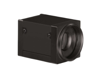
AcquCAM 5G

Down load Catalog – ACQUCAM 5G

  • USB 3.0 Color Camera
  • 2/3 ” Pregius Sensor
  • 2448 x 2048 pixel
  • Trigger input and I/O
  • Acquisition of images with sufficient contrast
  • High Sensitivity Sensor
  • Fast, seamless image transfer
  • Available of images with a very wide field of view
  • Low Noise Sensor

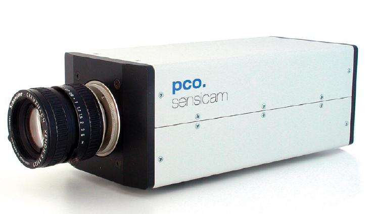
AcquCAM 5G color camera has a USB 3.0 interface and is the perfect solution for many industrial automation, quality assurance, security, surveillance and medical applications. The color camera ships with the very sensitive 2/3 inch CMOS Pregius  sensor. With up to 38 images per second, the AcuqCAM 5G is a low cost, yet highly versatile imaging solution. The camera includes a C to CS mount adapter, making it compatible to C and CS mount lenses. Using the optional CS to M12 board lens adapter, the camera is
also compatible to M12 board lenses.

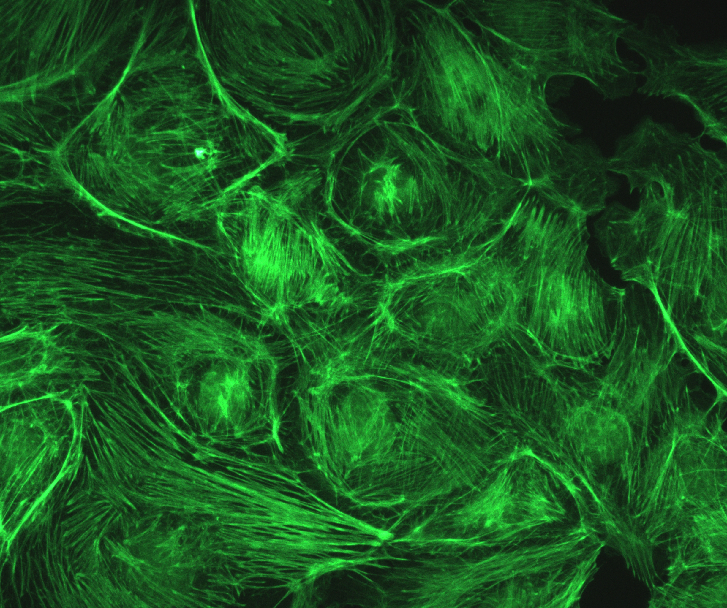
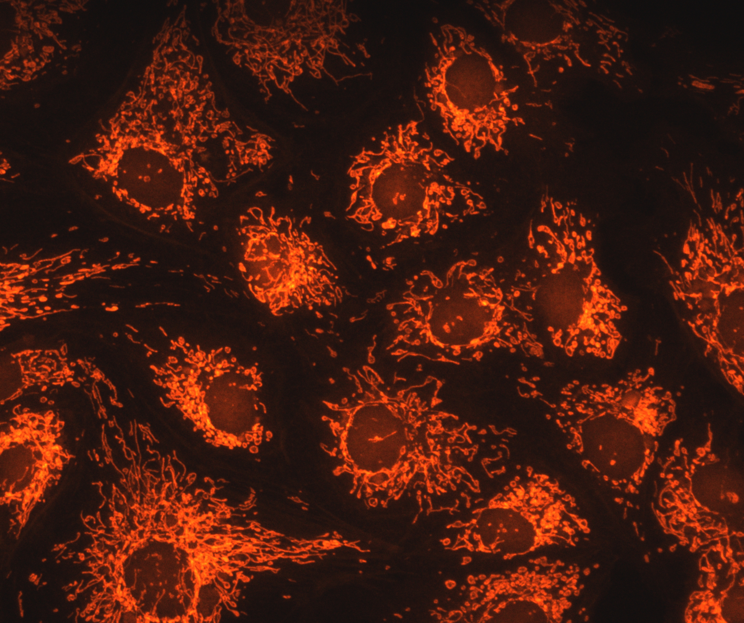
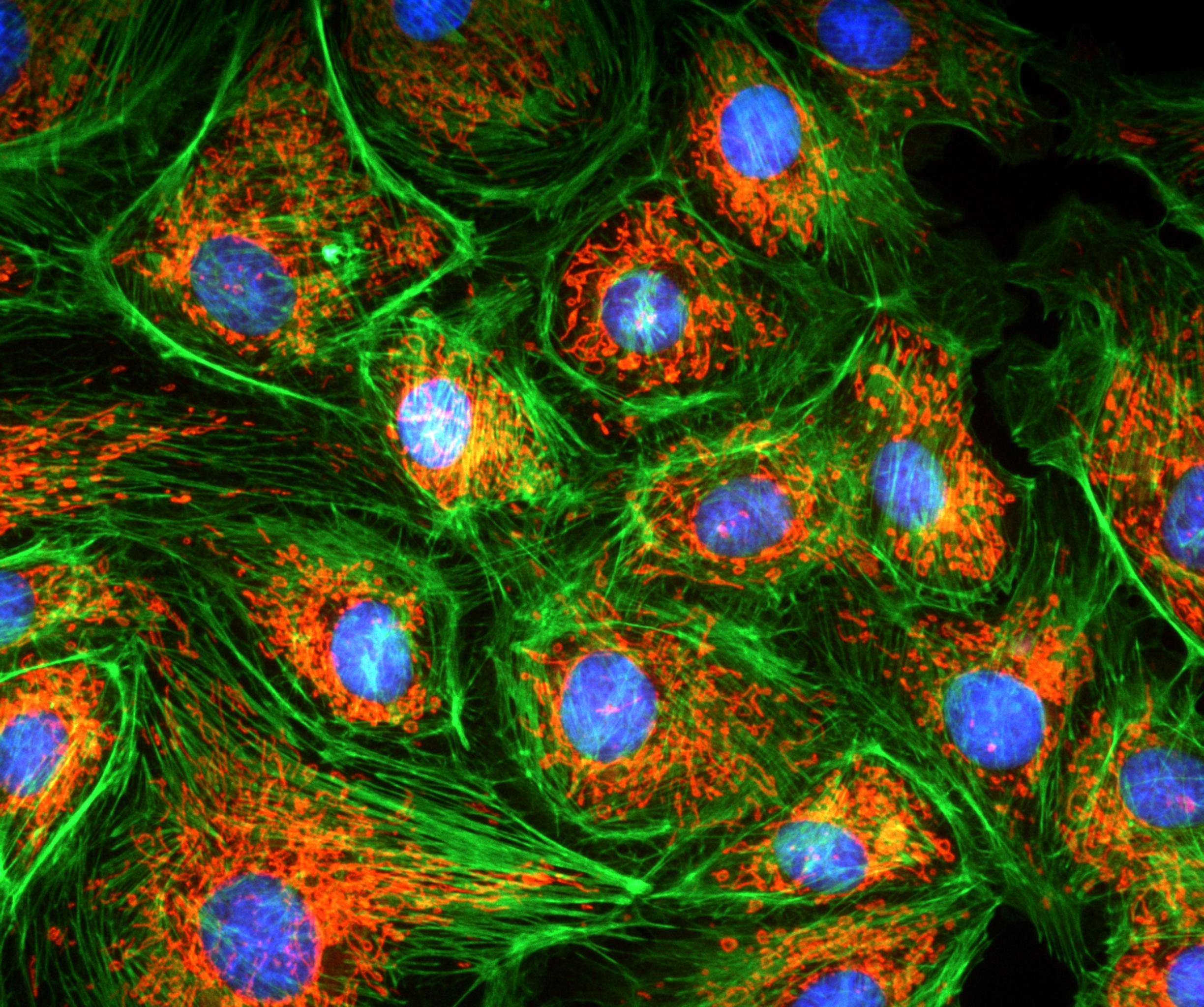
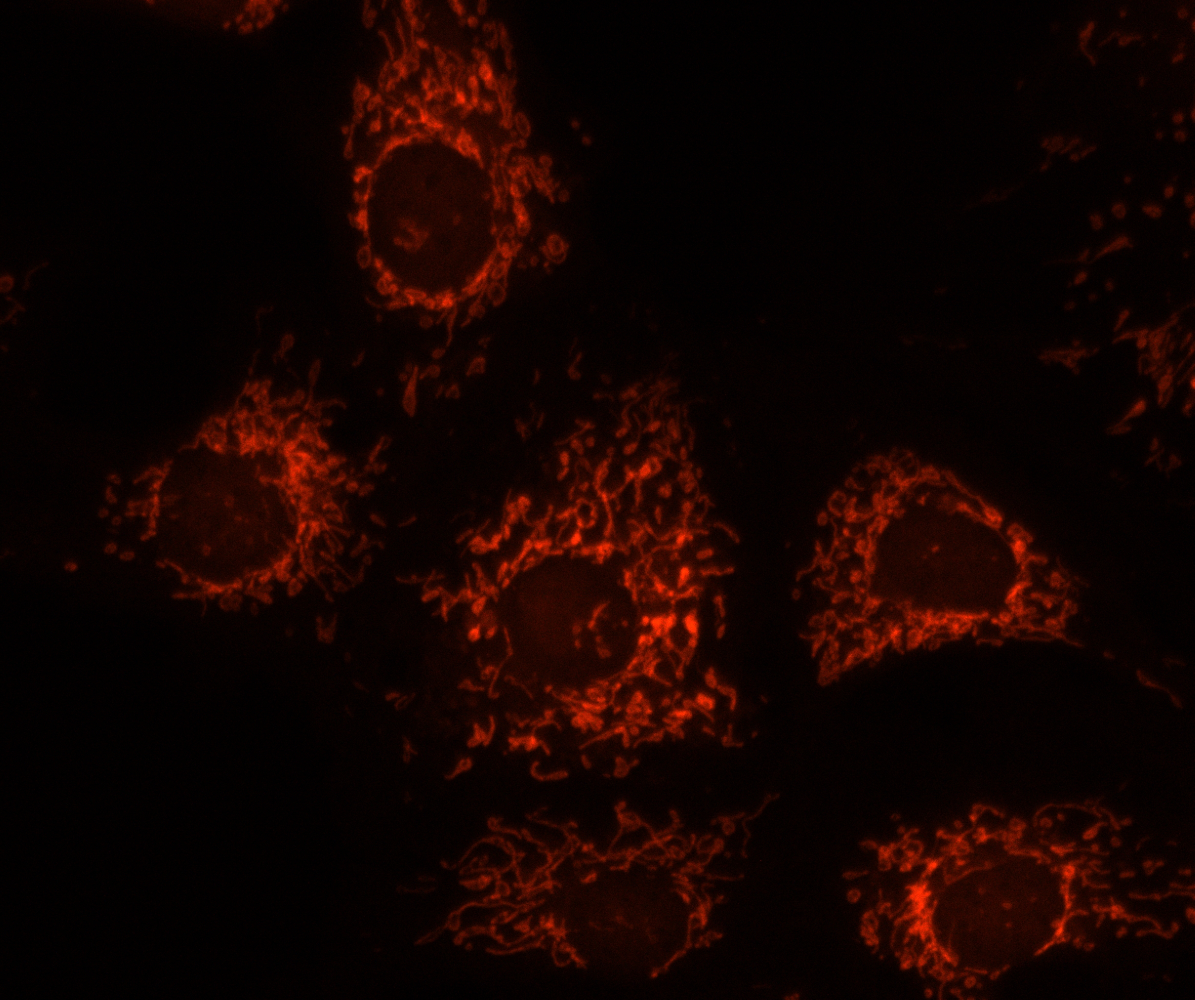
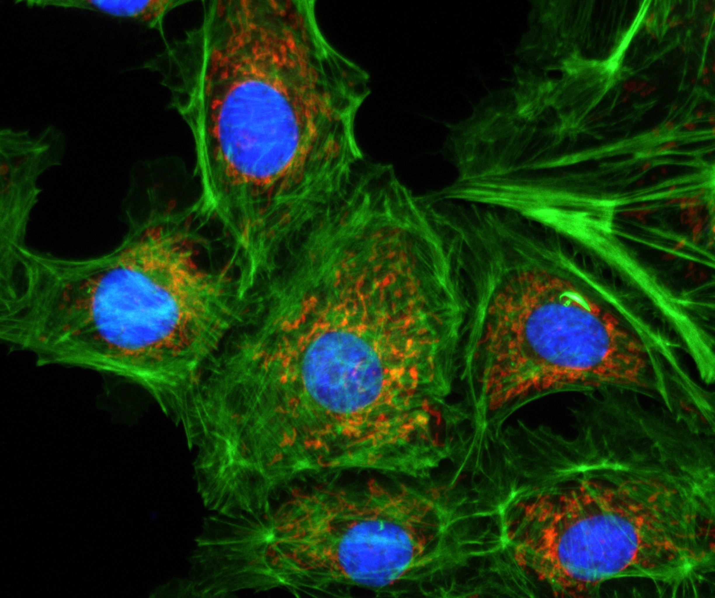
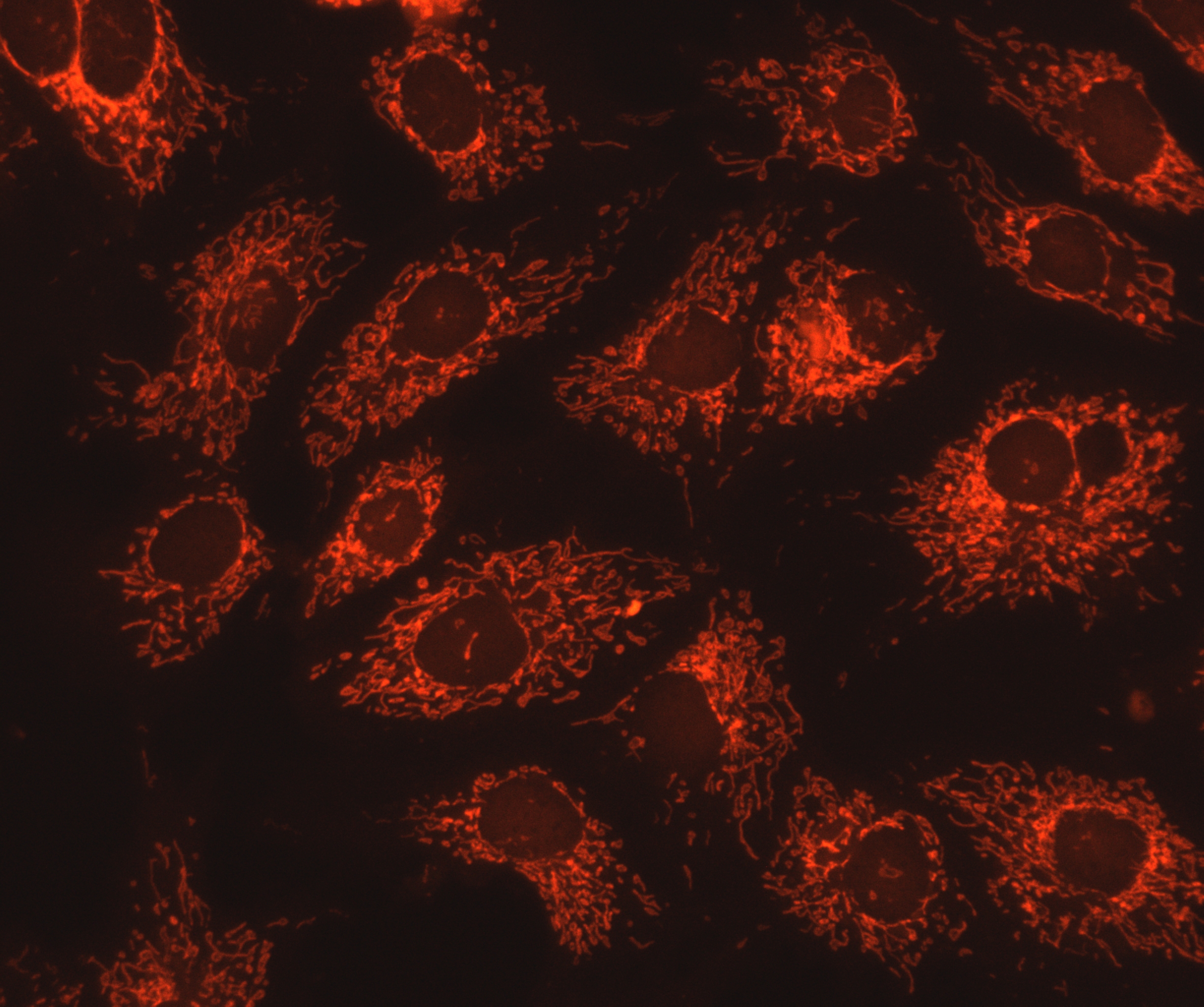
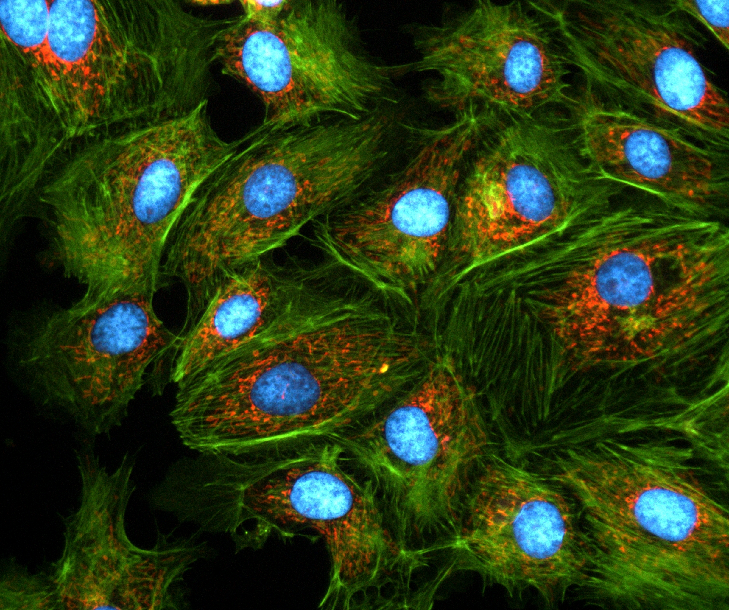
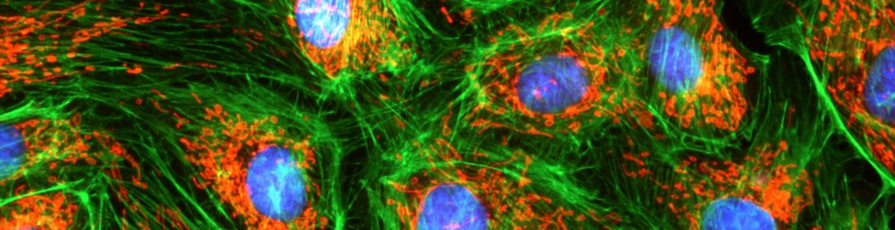
Images with AcquCAM 5G
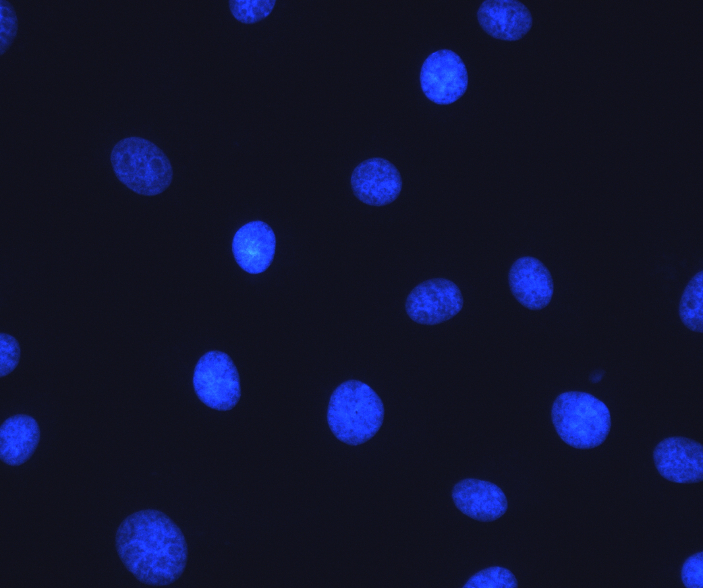
Sample : bovine pulmonary artery endothelial cell, BPAEC
Filter Set : JNO-U(B), B(B), G(B)
Camera : AcquCAM 5G
C-mount adapter : 1X
Lens : UPLNAPO40X
Above three images are merged and processed using ARM S/W
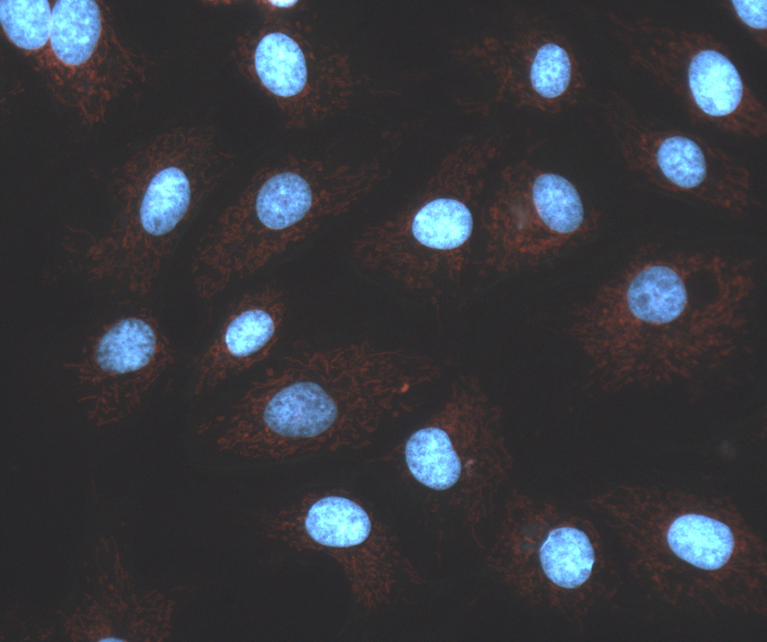
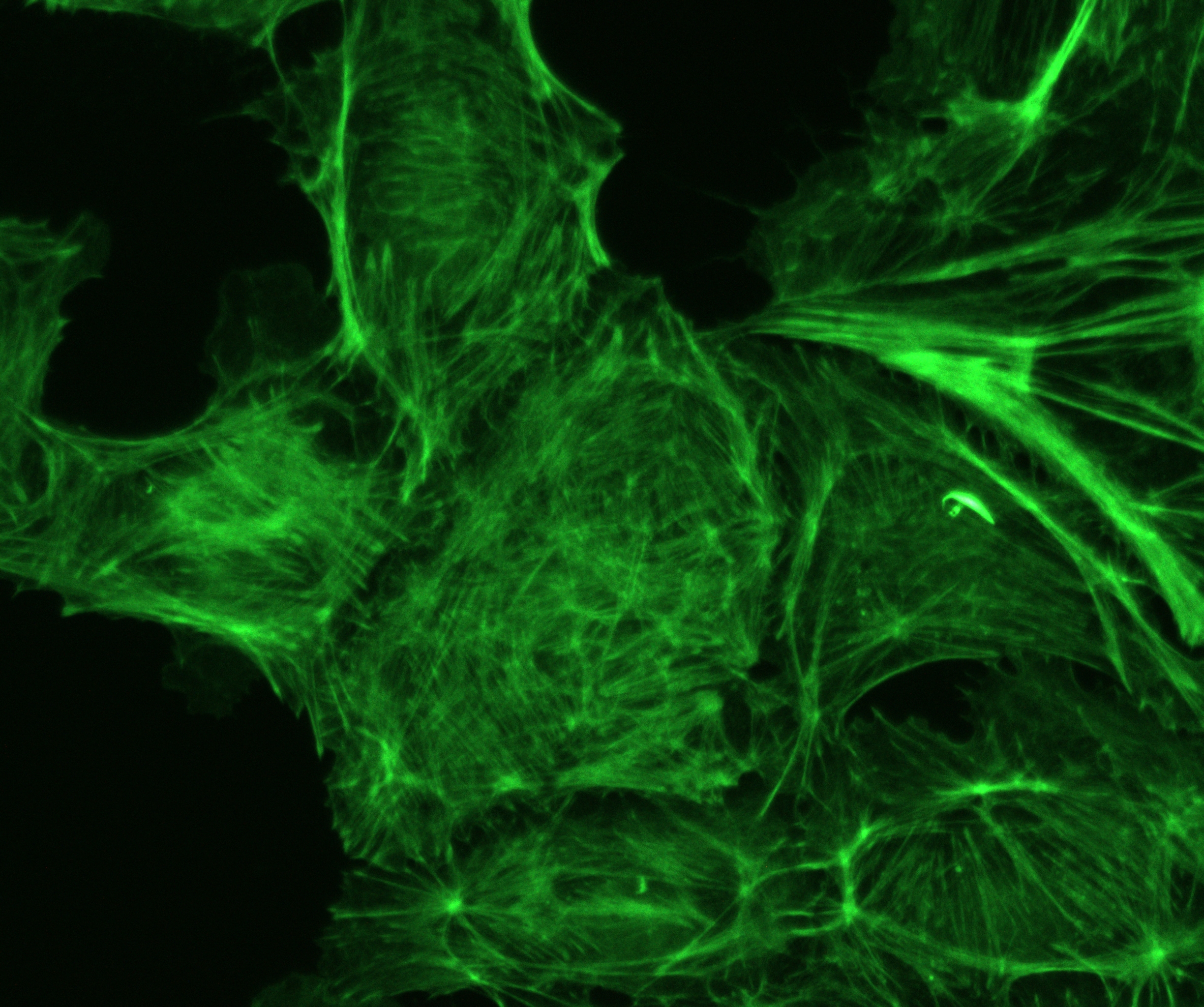
Sample : bovine pulmonary artery endothelial cell, BPAEC
Filter Set : JNO-U(B), B(B), G(B)
Camera : AcquCAM 5G
C-mount adapter : 1X
Lens : PlanApo60xWLSM
Above three images are merged and processed using ARM S/W
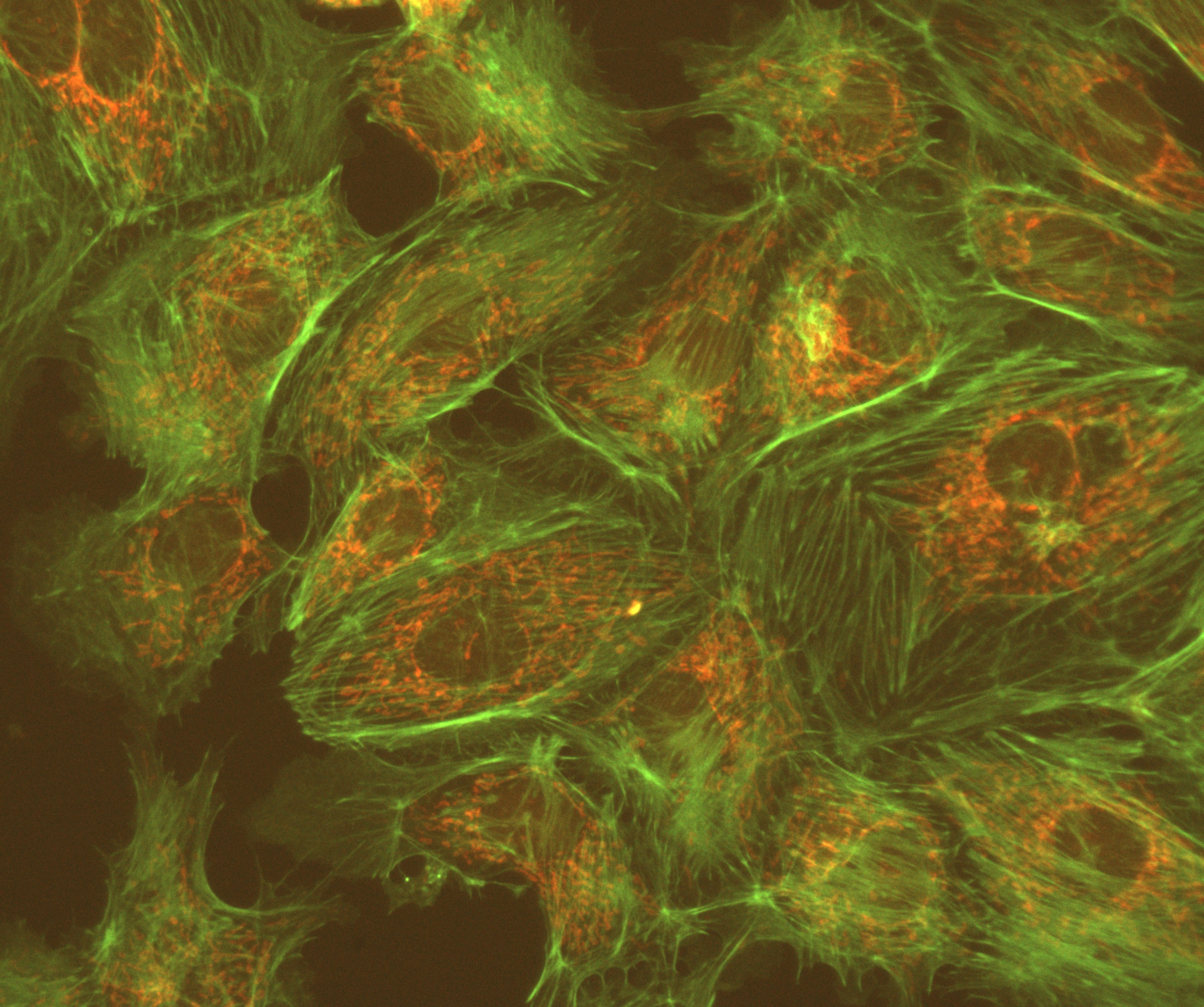
Sample : bovine pulmonary artery endothelial cell, BPAEC
Filter Set : JNO-U(L), B(L), G(L)
Camera : AcquCAM 5G
C-mount adapter : 1X
Lens : UPLNAPO40X
Above three images are merged and processed using ARM S/W

Specification

SpecDescription
Size2/3 inch
ResolutionH: 2448, V: 2048
Pixel sizeH: 3.45 µm, V: 3.45 µm
Video formats& Frame rate2448×2048 RGB64 @ 38 fps
2448×2048 RGB32 @ 35 fps
2448×2048 Y16 @ 37 fps                             2448×2048 Y411 @ 38 fps
2448×2048 YUY2 @ 37 fps
Dynamic range8 / 12 bit
Exposure time25 ㎲ to 4s
Lens mountC/Cs mount
InterfaceUSB 3.0
Power supply4.5 to 5.5 VDC
DimensionsH: 29 mm, W: 29 mm, L: 43 mm
Mass65g

ARM

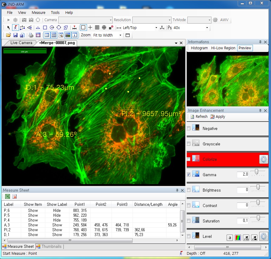
ARM is image analysis software for JNOPTIC AcquCAM cameras. This S/W is interchangeable with all WDM cameras regardless of camera brand and model and even more the most strength point is simple and easy use of length, area, angle, etc.
-> For more details, click here.

More images with AcquCAM 5G

-> For more details, click here.

TOMORO Frame Grabber 2

TOMORO Frame Grabber 2

이제 영상 분석을 위해 더 이상 고가의 Frame Grabber를 사용하지 마십시오.

TOMORO Frame Grabber 2 는 기본에 충실한 합리적인 가격의 Frame Grabber 입니다.

기존 TOMORO Frame Grabber 1에 비해 화질 및 성능을 개선되었으며 안정성 역시 향상 되었습니다.

TOMORO Frame Grabber 2는 절전 모드 및 Hibernation 후에도 끊김 없이 Live 영상을 출력하며 동영상 저장도 지원합니다.

주의 사항

TOMORO FRAME GRABBER2는 INTEL 의 메인보드의 칩셋을 100% 지원합니다.
타 업체의 칩셋과의 호환성을 먼저 체크 해 주시길 바랍니다.
(현재 지원하지 않는 칩셋 : SIS 963Lua)

Specifications

Input Connector Composite video (BNC) 1 EA
S-Video Color input connector (4-pin DIN) 1 EA
RCA connectors 2EA
Video Decoder NTSC : 640×480 (12.27MHz)
PAL : 768×576 (14.75MHz)
Video Format 24/16/15 bit RGB or YUV 4:2:2
Y8 monochrome
Bundle Software File format : JPG, BMP
Full Screen Display
Cross bar display on Live image
Time-lapse capture
Live movie capture
TWAIN interface
Dimensions (W x H x D) 120 x 140 x 20 mm
Mass Approx ~100g
Interface Standard PCI slot

Intensified Retiga

Intensified Retiga


저조도 및 나노초 (nanosecond) 노출의 고속 이미징 어플리케이션 요구를 위한 GEN III 이미지  intensifier


Features

18 mm Gen III Intensifier

Gate pulse from 50 ns to 17.9min

Intensifier Luminous Gain up to 80,000

Independent trigger and gate control

1360×1030 pixels

6.45 µm x 6.45 µm

Binning up to 8×8


Applications

Low Excitation Fluorescence Microscopy

Chemiluminescence

Bioluminescence

Single Molecule Fluorescence

Flow Analysis

Stop Action Imaging


Models

Intensified Retiga Extended Blue
Model: INT-RET-F-BLUE

Intensified Retiga Standard
Model: INT-RET-F


Retiga Exi

Retiga Exi


생명과학과 산업 분야의 극저조도 / brightfield /darkfield / fluorescence / NIR 이미징을 위한 고감도 정량 분석용


Features

1392×1040 pixels

6.45 µm x 6.45 µm

2/3″ optical format

Sensor Readout Noise 8e-

Dark Current 0.15e-/ pix/s cooled

Electronic Shutter

Binning up to 8×8

ROI

Cooling 25°C below ambient

External trigger


Applications

Brightfield/Dark Field Microscopy

Fluorescence Microscopy

Pathology, Histology, Cytology

GFP, FISH

DNA analysis

NIR Imaging

Motility and Motion Analysis

Semiconductor Inspection

Failure Analysis

Forensic Analysis


Models

Retiga EXi Fast 1394 Mono Cooled
Model: RET-EXi-F-M-12-C-IR
Model: RET-EXi-F-M-12-C

Retiga EXi Fast 1394 Color Cooled
Model: RET-EXi-F-CLR-12-C

Retiga EXi Fast 1394 Mono
Model: RET-EXi-F-M-12-IR
Model: RET-EXi-F-M-12

Retiga EXi Fast 1394 Color
Model: RET-EXi-F-CLR-12

Retiga EXi Mono Cooled
Model: RET-EXi-M-12-C-IR
Model: RET-EXi-M-12-C

Retiga EXi Color Cooled
Model: RET-EXi-CLR-12-C

Retiga EXi Mono
Model: RET-EXi-M-12-IR
Model: RET-EXi-M-12

Retiga EXi Color
Model: RET-EXi-CLR-12


Retiga EXi Fast 1394 Data Sheet

Retiga EXi Data Sheet

Retiga 4000R

Retiga 4000R


생명 과학 및 산업 분야의 저 조도 / birightfield/ darkfield/ flourescence 이미징을 위한 고정밀 정량 분석용


Features

Kodak Sensor KAI-4021

2048 x 2048 pixels

7.4 µm × 7.4 µm pixel size

Linear Full Well 40,000e-

QE 55% at 500nm

Electronic Shutter

Binning up to 8 × 8

ROI

Cooling 25°C below ambient

External trigger


Applications

Brightfield, Phase-Contrast & Dark Field Microscopy

Fluorescence Imaging

Pathology, Histology, Cytology

DNA analysis

Metallurgical Microscopy

LCD Inspection

Manufacturing Quality Control

Failure Analysis

Forensic Analysis

Automated Imaging


Models

Retiga 4000R Fast 1394 Mono Cooled
Model: RET-4000R-F-M-12-C

Retiga 4000R Fast 1394 Mono
Model: RET-4000R-F-M-12

Retiga 4000R Fast 1394 Colour Cooled
Model: RET-4000R-F-CLR-12-C

Retiga 4000R Fast 1394 Colour
Model: RET-4000R-F-CLR-12


Retiga 2000R

Retiga 2000R


생명 과학 및 산업 분야의 저 조도 / birightfield/ darkfield/ flourescence 이미징을 위한 고정밀 정량 분석용


Features

Kodak Sensor KAI-2020

1600 x 1200 pixels

7.4 µm × 7.4 µm pixel size

Linear Full Well 40,000e-

QE 55% at 500nm

Electronic Shutter

Binning up to 8 × 8

ROI

Cooling 25°C below ambient

External trigger


Applications

Brightfield /Dark Field Microscopy

Fluorescence Imaging

Pathology, Histology, Cytology

FGP, FISH

DNA analysis

NIR Imaging

Motility and Motion Analysis

Semiconductor Inspecition

Failure Analysis

Forensic Analysis


Models

Retiga 2000R Fast 1394 Mono Cooled
Model: RET-2000R-F-M-12-C

Retiga 2000R Fast 1394 Mono
Model: RET-2000R-F-M-12

Retiga 2000R Fast 1394 Colour Cooled
Model: RET-2000R-F-CLR-12-C

Retiga 2000R Fast 1394 Colour
Model: RET-2000R-F-CLR-12

Dicam pro

Dicam pro

intensified digital 12bit CCD camera system


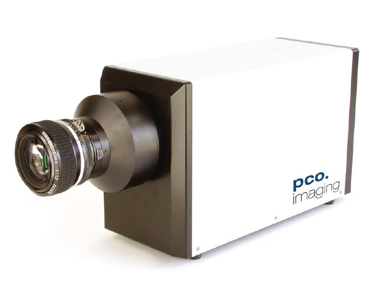
PCO사에서 제작한 Dicam pro 카메라는 고속의 intensified CCD를 장착한 시스템입니다.

 12bit 이미지에 130만화소 고해상도를 지원하며 S/N 비가 낮고 단일 원자 검출 능력이 뛰어난 것이 특징입니다.

 카메라와 PC는 고속의 직렬 광데이터 링크로 연결됩니다.

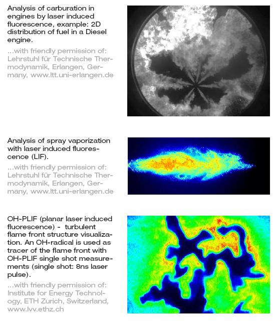
Dicam pro의 intensified digial CCD 카메라 시스템은 spray imaging, laser induced fluorescence imaging or ballistics와 같이 초고속의 카메라 application을 요구로 하는 분야에 완벽하게 적합합니다.


■ Features

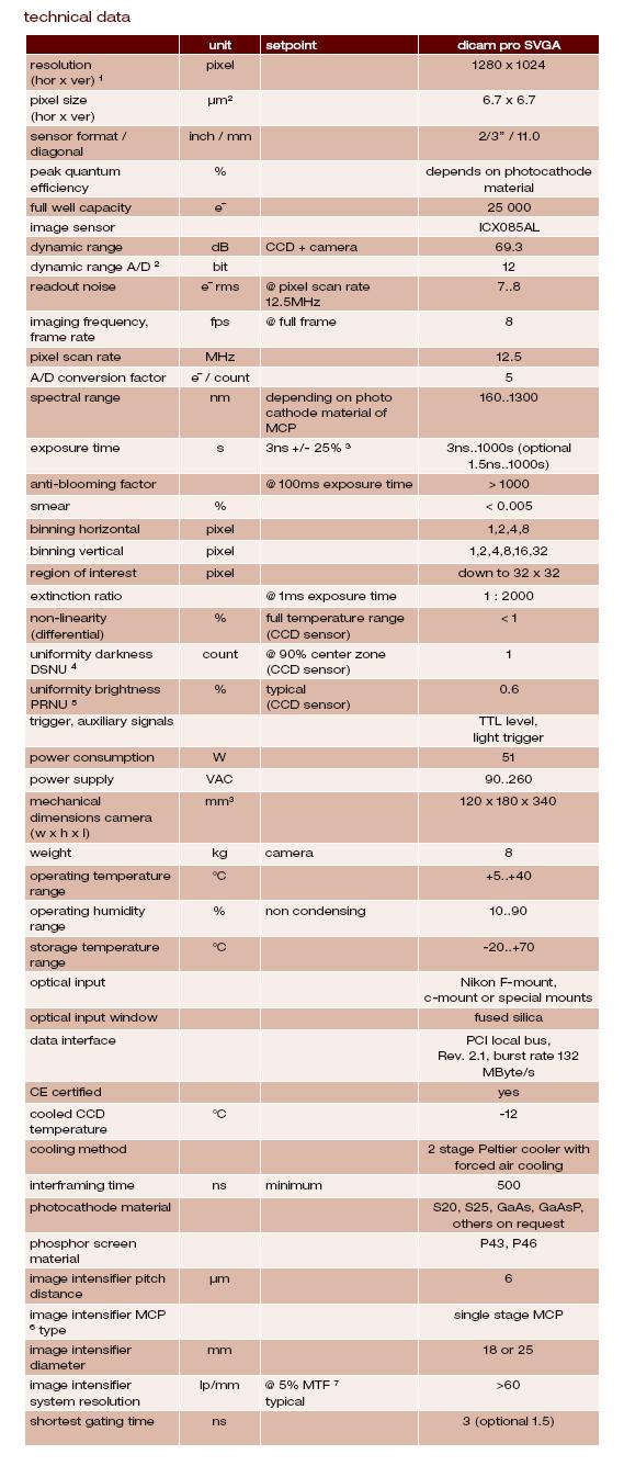
  ▲ fast shutter down to 3ns (optional 1.5ns)

  ▲ excellent sensitivity of the system allows single photon detection

  ▲ 12bit dynamic range

  ▲ high resolution MCP-image intensifier & CCD (1280x1024pixel)

  ▲ exposure times from 3ns (optional 1.5ns) – 1000s

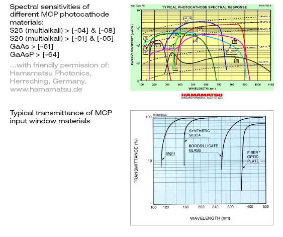
  ▲ spectral sensitivity from UV to NIR

  ▲ PCI interface board “plug & play

  ▲ binning (horizontal & vertical)

  ▲ thermo-electrical cooling (Peltier) of CCD image sensor down to -12°C

  ▲ optical or electrical triggering

  ▲ various MCP photocathodes – multialkali, S20, GaAs, GaAsP

  ▲ two discrete images with an interframing time of 500ns (PIV)

  ▲ multiple exposures

  ▲ serial high speed data transfer via fiber optic link (FOL)

  ▲ free software camware and software development kit included


■ Spectral response of MCP


■ Specification


■ Examples of applications

Pixelfly QE

Pixelfly QE

high performance digital 12bit CCD camera system


PCO사에서 제작한 Pixelfly QE 카메라는 12bit cooled CCD가 장착된 초소형 고성능 이미징 시스템입니다.

 공간을 차지하는 열전자 cooling 장치가 없이도 디지털 온도 보정이 가능하여 카메라를 소형화 하였습니다..

 이 시스템은 standard PCI나 compact PCI보드와 고속 직렬 데이터 링크를 통해 연결되는 초소형 카메라 헤드로 구성되어 있습니다.

Pixelfly의 디지털 카메라 시스템은 microscopy, spectroscopy, quality control과 같이 여러 과학 및 산업 이미지 application 분야에 완벽하게 적합합니다.


■ Features

▲ superior quantum efficiency up to 65%

  ▲ ultra compact design

  ▲ 12bit dynamic range

  ▲ high resolution (1392 x 1024pixel)

  ▲ temperature compensated

  ▲ exposure times from 5μs – 65s

  ▲ readout noise typ. 7e- rms

  ▲ serial high speed data transfer up to 10m

  ▲ standard PCI or compact PCI control board

  ▲ integrated front-end processor with opto-coupler input and highside driver

  ▲ free software camware and software development kit included


■ Quantum Efficiency


■ Specification


■ Examples of applications