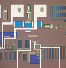
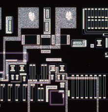
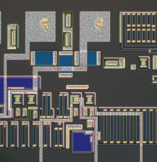
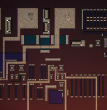
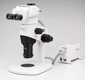
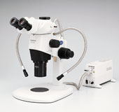
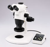
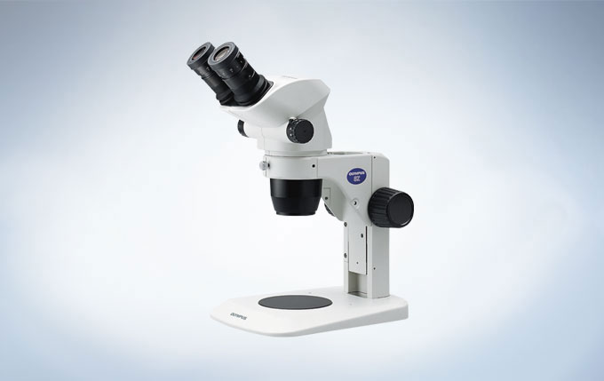
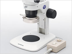
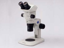
SZ61과 SZ51의 새로운 Comfort View 접안렌즈는 범용 LED 조명 스탠드와 함께 사용되어 LED 기술의 모든 이점을 제공함과 동시에 동공 수차 제어를 통해 빠르고 편안한 관찰을 보장합니다. Greenough 광학 시스템의 영상 형성 경로의 낮은 집중각은 우수한 이미지 평탄도와 초점 심도를 보장합니다. 최상급 광학 코팅은 높은 색 재현성(Color fidelity)을 제공합니다. 정전기 방지 물질과 코팅은 표본을 정전기 방전으로부터 보호합니다.SZ61 Zoom 실체 현미경은 일상 혹은 심화 현미경 관찰, 특히 디지털 이미징 및 문서화가 필요한 경우에 이상적입니다.경제적인 SZ51 Zoom 실체 현미경은 생명 과학을 위한 폭넓은 범위의 기능들을 제공합니다.
Compact Stereo with High-Quality Optics
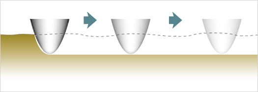
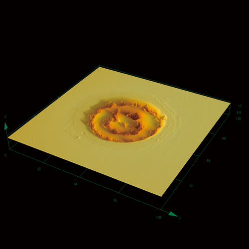
Greenough Optical System
The 10-degree angle of convergence in the Greenough optical system secures excellent image flatness with large depth of field. The careful selection of lens surface coatings and glass materials in the entire optical system makes it possible to observe and document specimen in their original, authentic colors. The V-shape optical path ensures a slim zoom body – ideal for integration into other equipment or standalone use.
그리너프(Greenough) 광학 시스템의 광로 도면
Wide Zoom Ratio
The SZ61’s class-leading magnification range extends from 6.7x to 40x (using 10x eyepieces), with a wide zoom ratio of 6.7:1, enabling smooth, macro-to-micro zooming that speeds routine workflows. The SZ51 provides the magnification rage of 8x to 40x (using 10x eyepieces), with a wide zoom ratio of 5:1.
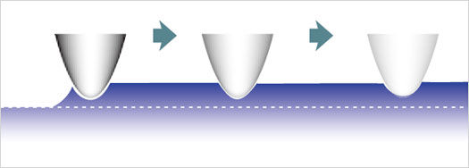
3 Dimensional Viewing
The optimum inward angle allows just the right combination of high level flatness and depth of focus for 3D viewing. Even a specimen with significant depth can be brought into focus from top to bottom for faster inspection.
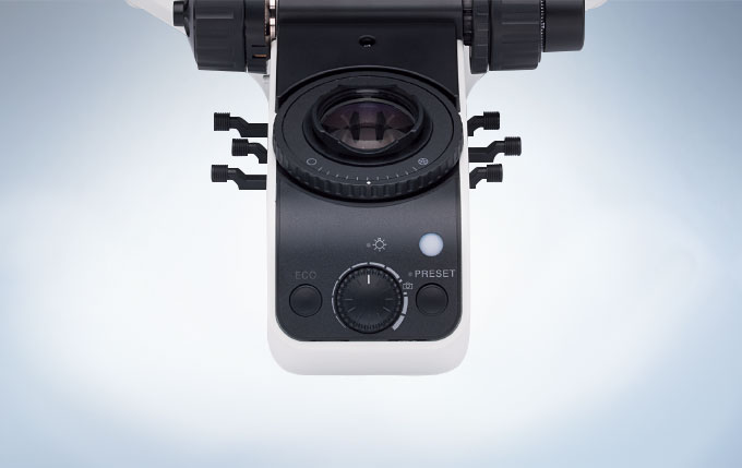
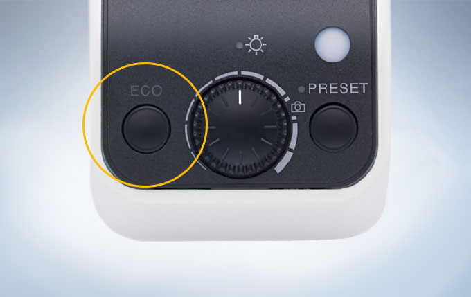
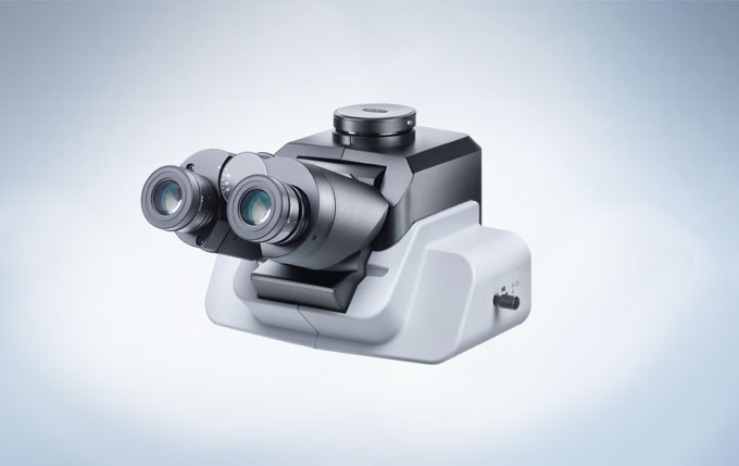
Comfortable and Reliable Design for Routine Work
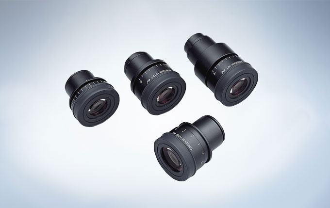
ComfortView Eyepieces for Reduced Eyestrain
Quick, comfortable observation is ensured by this exclusive eyepiece design featuring pupil aberration control and appropriate positioning of the eyepoint. Also, superior optical coatings render true color images with fine detail.
컴포트뷰 접안렌즈
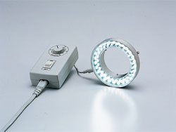
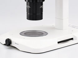
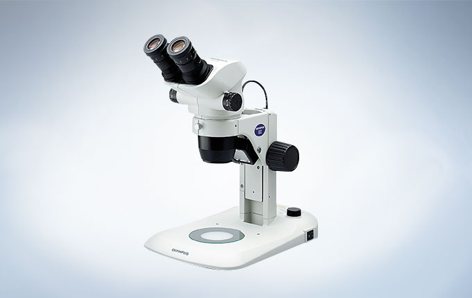
LED Illumination
The universal reflected/transmitted light LED illumination stand brings together all the advantages of LED technology. The flat, high-brightness LEDs allow successful integration of transmitted illumination into a very slim base, which in turn facilitates easy specimen access and manipulation.
LED 조명
Electrostatic Discharge Protection
The main body and major accessories can quickly eliminate static electricity with the use of antistatic materials and coatings. This prevents a specimen under observation from electrostatic damage.
Choice of High-Performance Bodies
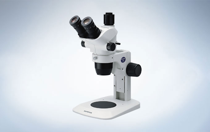
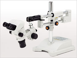
The SZ61 and SZ51 zoom bodies provides 2 different magnification ranges, and each one is ergonomically designed with a 45-degree inclination tube as the standard model. For special applications where the zoom body has to be tilted of use with other equipment of mounting on a universal stand, the built-in models with 60-degree inclination tube are available. For documentation purpose, a trinocular model is available for the quick and easy attachment of digital cameras.
With the Olympus LEXT OLS4100 laser scanning digital microscope non-contact 3D observations and measurements of surface features at 10 nanometer resolutions are easy to produce. The OLS4100 industrial microscope has distinctive features for fast image acquisition and high-resolution microscope images over a wider area.
뛰어난 측정 성능
세계 최초* 두개의 성능 보증
고품질 이미지
더 쉽게, 더 빠르게, 더 광범위하게.
뛰어난 측정 성능
넓은 샘플 범위
85°이상의 경사도 이미지
OLS4100은 높은 N.A값을 갖는 전용 대물 렌즈와 405nm 레이저의 성능을 극대화 시킨 광학 시스템에 의해서 기존에 측정 불가능했던 급경사면의 이미지를 손쉽게 취득할 수 있습니다.
수차를 최소화한 전용렌즈
10nm의 높이 분해능을 가진 마이크로 프로파일 측정
(대물 렌즈 : MPLAPON50XLEXT) STEP 높이 표준 B 타입, PTB-5, 독일, 마이크로 전자 연구소 6nm 단차를 감지
405nm의 단파장 레이저 빛과 높은 N.A의 전용 대물 렌즈를 사용하여 최대 0.12μm의 평면 분해능을 실현. 샘플 표면의 서브 마이크론 측정이 가능합니다. 또한 고정밀 리니어 스케일과 IZ 커브를 이용한 CFO 검색 (23 페이지 참조)를 채택하여 10nm 이하의 높이 차이를 감지 할 수 있습니다.
반사율 차이에도 대응
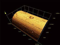
다이아몬드 전기 공구 대물 렌즈 : MPlanApoN50xLEXT
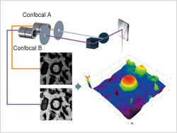
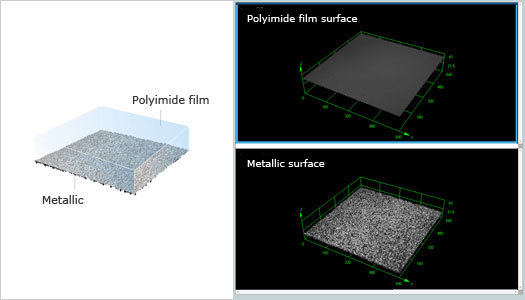
OLS4100은 2개의 공초첨 광학 시스템을 탑재한 듀얼 컨포칼 시스템을 사용합니다. 고감도 디텍터(검출기)와 결합하여 서로 다른 반사율을 갖는 샘플에서도 선명한 이미지 구현이 가능합니다.
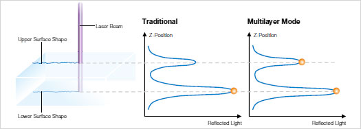
멀티 레이어기능으로 투명한 물체의 측정에 대응
멀티 레이어 모드 OLS4100에 추가 된 멀티 레이어 기능은 여러 레이어 (층)의 반사광 강도의 피크를 인식하여 각 층의 두께를 측정할 수 있습니다.
투명 소재의 멀티 레이어 관찰/측정 멀티 레이어 기능을 사용하면 투명 샘플의 윗 표면에 있는 투명 필름의 관찰과 측정이 가능합니다. 레진이 코팅 되어 있는 샘플에서도 투명체 각 층의 거칠기와 두께의 측정도 가능합니다.
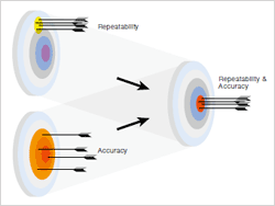
세계 최초* 두개의 성능 보증
정확도와 반복성
측정 기기로서의 성능은 두 개의 다른 용어로 표현됩니다. 그것은 ‘정확도’과 ‘반복성’입니다. “정확도”는 참값에 얼마나 접근하고 있는지를 나타내고 “반복성”은 여러 번 측정에서 얼마나 변동이 적은가 여부를 나타내는 것입니다. OLS4100은 레이저 현미경으로 업계 최초로 *이 두 성능을 보증합니다.
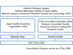
보증 체계 시스템
OLS4100은 모든 부품이 엄격한 시스템 아래 하에 제조되고 있습니다. 대물 렌즈부터 본체까지 자사 공장에서 일관 생산하고 엄격한 검사 기준을 거쳐 출하됩니다. 최종 조정과 교정은 실제로 사용되는 환경에서 전문 기술자가 수행합니다.
넓은 범위의 측정 타입
단차 측정
이 모드는 단면 프로파일에서 임의의 두 점 사이의 단차를 측정 할 수 있습니다.
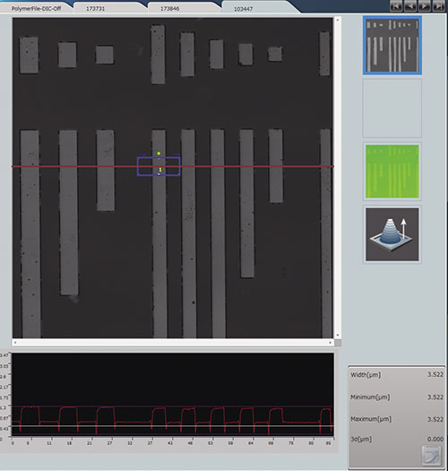
표면 거칠기 측정
이 모드는 한 라인의 선 거칠기, 표면 전체의 거칠기 측정이 가능합니다.
면적/부피 측정
단면 프로파일에서 임의의 임계 값을 설정하여 그 상부 또는 하부의 체적을 측정 할 수 있습니다.
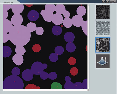
입자 측정(옵션)
이 모드는 임계 값 레벨의 설정, 그리고 관심 영역 내에서 감지 범위의 설정 및 분리 기능을 가진 입자의 자동 분리를 할 수 있습니다.
기하학적 측정
이 모드는 이미지상의 임의의 두 점 사이의 거리를 측정 할 수 있습니다. 원형, 사각형 등의 기하학적 모양과 각도를 측정합니다.
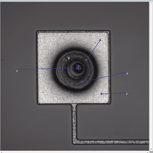
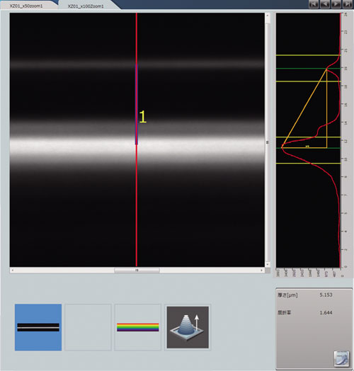
필름 두께 측정(옵션)
이 모드는 굴절율의 변화를 감지하여 투명 필름의 두께를 측정 할 수 있습니다.
자동 에지 검출/측정(옵션)
이미지의 가장자리를 자동으로 감지하여 선폭 · 원의 측정을 할 수 있습니다. 사용자에 의한 측정 오차를 줄일 수 있습니다.
OLYMPUS Stream (옵션)
향상된 이미지 분석 성능을 위한 업무개선 솔루션 소프트웨어 “Olympus Stream”(옵션)에서는 입자 크기 분석 및 비금속 함유율 등이 가능하고 OLS4100으로 부터 바로 연동이 가능합니다.
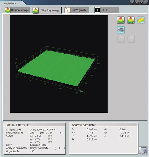
더욱 더 진화된 거칠기 측정
LEXT OLS4100 파라미터
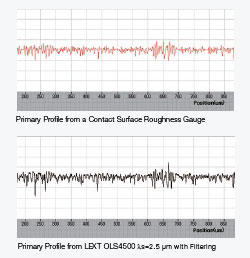
OLS4100은 표면 거칠기 측정기의 새로운 기준을 목표로 개발되었습니다. 필요한 대부분의 거칠기 파라미터 필터를 보유하고 있습니다. 그렇기 때문에 기존의 접촉식 표면 거칠기 측정기를 사용하는 사용자는 기존의 장비와 일치한 출력 결과를 얻을 수 있습니다.. 또한 OLS4100은 거칠기 전용 모드가 있어 자동 라인 스티치로 최대 100mm까지 거칠기 측정이 가능합니다. OLS4100은 접촉식 표면 거칠기 측정기와 같은 거칠기 (2 차원) 파라미터를 보유하고 있습니다. 접촉식 표면 거칠기 측정기와 같은 조작성, 호환 측정 결과를 얻을 수 있습니다.
차세대 파라미터에 대응 OLS4100은 ISO25178 규격 거칠기 (3 차원) 파라미터를 보유하고 있습니다. 평면 영역에서 평가를 실시하는 것으로, 높은 신뢰성이 있는 분석이 가능합니다.
진폭 파라미터
Sq, Ssk, Sku, Sp, Sv, Sz, Sa
기능 파라미터
Smr(c), Sdc(mr), Sk, Spk, Svk, SMr1, SMr2, Sxp
체적 파라미터
Vv(p), Vvv, Vvc, Vm(p), Vmp, Vmc
평면 파라미터
Sal, Str
LEXT OLS4100은 표면 거칠기 측정기의 결과와 호환성을 가지고 있습니다.
마이크로 거칠기
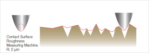
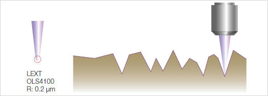
접촉식 표면 거칠기 측정기는 스타일러스의 팁 지름보다 작은 마이크로 표면을 측정 할 수 없습니다. 레이저 현미경은 레이저 스폿 직경이 미세하기 때문에 미세형상을 높은 분해능으로 거칠기 측정 할 수 있습니다.
비 접촉 측정
접촉식 표면 거칠기 측정기는 스타일러스에 의해 부드러운 샘플의 표면을 긁어 손상시키거나 변형시킬 가능성이 높습니다. 또착 접착성이 있는 샘플에서는 스타일러스에 끌려 정확한 측정 결과가 나오지 않습니다. 레이저 현미경은 비접촉이기 때문에 표면 상태에 영향을 받지 않고 정확한 거칠기 측정을 할 수 있습니다.
부드러운 샘플접착성 샘플
마이크로 영역에서의 측정
접촉식 표면 거칠기 측정기는 스타일러스를 표면에 접촉하지 않고는 측정 할 수 없습니다. 레이저 현미경은 위치를 정확하게 확인하고, 목표한 마이크로 영역에서 거칠기 측정을 아주 쉽게 할 수 있습니다.
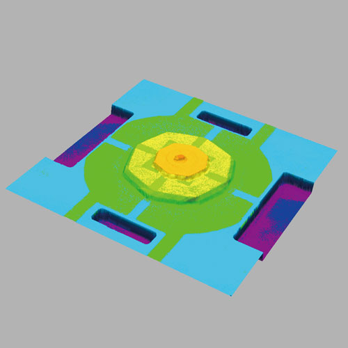
고품질 이미지
선명한 3D 컬러 이미지
통합이미지의 3가지 유형
LEXT OLS4100은 동일한 시각의 컬러 이미지, 레이저 현미경이미지, 높이 이미지를 동시에 얻을 수 있으며, 각각 2D・3D로 볼 수 있습니다. 레이저 현미경 이미지 뿐만 아니라 컬러 이미지도 초점이 맞는 이미지만 캡처 하기 때문에 선명한 이미지를 얻을 수 있습니다.
리얼 컬러 3D 이미지
공초점 3D 레이저 이미지
높이 정보
자연 컬러 재연
OLS4100은 백색 LED를 사용하며, 색 재현이 뛰어난 촬상 소자와 결합하여 선명하고 자연스러운 색조의 컬러 이미지를 생성 할 수 있습니다.
2D 컬러 이미지 (종이 위의 잉크젯 점, 대물렌즈 20x)
3D 컬러 이미지 (종이 위의 잉크젯 점, 대물렌즈 20x)
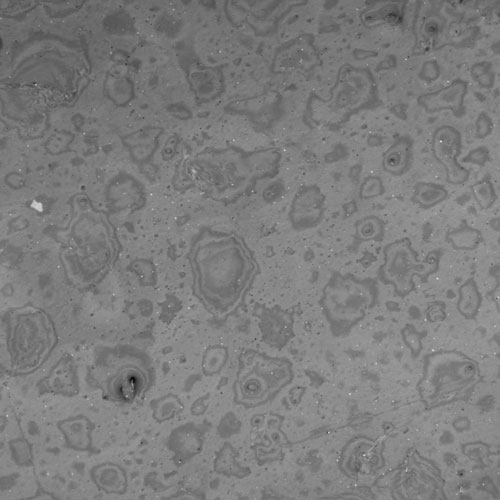
실제 표면 재현, 레이저 DIC (미분 간섭 대비)
미분 간섭 관찰(DIC)은 레이저 현미경의 분해능을 더 넘어선, 나노 미터 수준의 미세한 표면을 시각화하는 관찰 방법입니다. 이 레이저 DIC에 의해 OLS4100은 저배율의 라이브 관찰에서도 전자 현미경의 분해능과 같은 이미지를 얻을 수 있습니다.
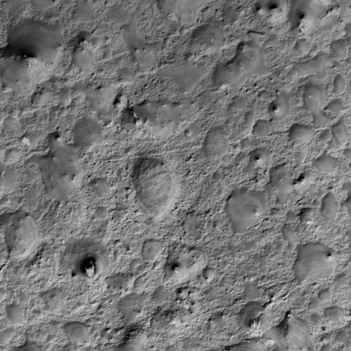
DIC가 없는 레이저 현미경 이미지(고분자 필름)
DIC가 있는 레이저 현미경 이미지(고분자 필름)
DIC가 없는 레이저 현미경 이미지(5x 대물 렌즈) STEP 높이 표준 타입 B, PTB-5, 독일 마이크로 전자 연구소
DIC가 있는 레이저 현미경 이미지(5x 대물 렌즈) STEP 높이 표준 타입 B, PTB-5, 독일 마이크로 전자 연구소
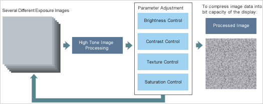
밝기와 대비의 최적 밸런스 HDR (High Dynamic Range) 이미지
OLS4100의 HDR기능은 다른 노출로 촬영 한 여러 가지 광학 현미경 이미지를 결합하고 개별적으로 밝기, 명암, 질감과 채도를 조정하여 넓은 동적 범위의 HDR 프로세스로 이미지화합니다. OLS4100은 대비가 부족한 샘플의 미세 형상도 선명히 라이브 화상으로 관찰 할 수 있습니다.
컬러 이미지 (고밀도 직물, 대물렌즈 20x, 줌 1x)HDR 컬러 이미지 (고밀도 구조, 대물렌즈 20x, 줌 1x)
알고리즘
측정 및 이미지 환경의 안전화
하이브리드 진동 완충 기구
OLS4100은 외부의 영향을 억제하여 측정 및 이미징 환경을 안정시키기 위해 본체에 코일 스프링과 댐핑 러버로 이루어진 ‘하이브리드 제진 장치 “를 내장하고 있습니다. 따라서 전용 제진대 없이도 측정이 가능합니다.
더 쉽게, 더 빠르게, 더 광범위하게.
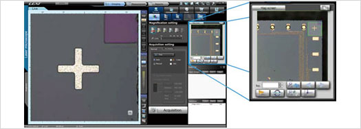
3단계의 간단 조작
OLS4100에서는 샘플을 스테이지에 올리는 것 만으로, 곧바로 관찰 · 측정을 시작할 수 있습니다. 간단한 이미지 획득, 측정, 보고서 작성 이 3 단계로 레이저 현미경에 익숙하지 않은 초보자도 쉽게 측정 방법을 마스터 할 수 있습니다.
항상 샘플의 위치를 알 수 있습니다.
매크로 맵 기능
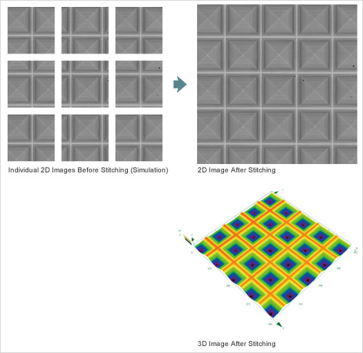
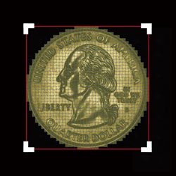
OLS4100에서는 매크로 맵 기능이 있기 때문에 “샘플이 지금 어디에 있는지”를 화면에 나타낼 수 있습니다. 전동 리볼버를 사용하기 때문에 저배율의 광범위한 샘플 이미지를 자동으로 표시합니다. 또한 스티칭 기능을 사용하면 기존에 비해 최대 441 배 넓은 시야의 매크로 맵을 만들 수있게 되었습니다.
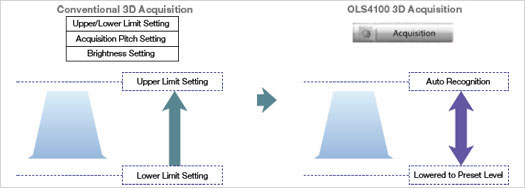
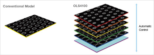
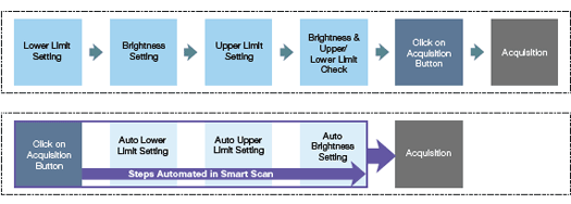
스마트 스캔 기능을 새롭게 첨가한 OLS4100. 기존의 3D 검사는 초보자에게는 어려운 복잡한 설정이 필요했지만, 스마트 스캔 기능을 통해 쉽게 3D 이미지 전송을 할 수있게 되었습니다. 상하단 설정 이외에 밝기 조정도 자동으로 수행되며 한 번의 클릭으로 이미지 전송이 가능하여 초보자도 숙련자와 같은 최적의 이미지 획득이 가능합니다.
자동 밝기 조절
평면 밝기 조절 높이의 범위 내에서 밝기 조절
획득 시간이 대폭 단축
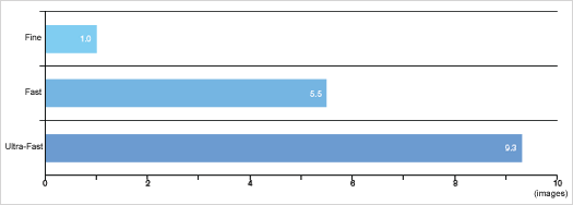
스캐닝 스피드의 고속화
새롭게 생긴 초고속 모드는 기존 고속 모드의 약 2배, 고화질 모드의 약 9배의 속도로 스캔 할 수 있습니다. 이를 통해 광범위의 Z축 방향의 획득과 고배율이 필요한 칼 끝 같은 급경사 각도를 가진 샘플의 측정도 단시간에 가능합니다. * 실제 스캔 시간은 Z축 방향의 범위 및 대물렌즈의 배율 등에 의해 변동됩니다.
동일 시간에 따른 이미지 획득 수:
실제 스캐닝 시간은 배율 과 Z축 획득 범위에 따라 달라집니다.
필요한 부분만을 고속 스캔
OLS4100 밴드 스캔 기능은 샘플 검사 범위를 한정함으로써 원래의 약 1 / 8의 고속 측정이 가능합니다.
풀 스캔을 한 획득
밴드 스캔을 한 획득(약 1/8 시간 단축)
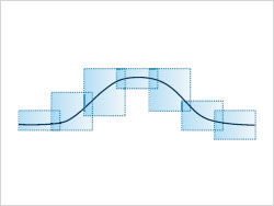
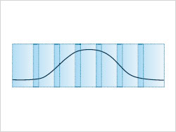
새로운 고속 스티칭 모드
넓은 영역의 스티칭 이미지에서 원하는 영역을 지정
매크로 맵과 같은 절차로 획득한 광범위의 이미지에서 3D 스티칭 범위를 지정할 수 있습니다. 최대 625장의 이미지 스티칭이 기존의 절반의 소요시간으로 생성 가능합니다. 스티칭의 범위를 지정하는 방법은 사각형뿐만 아니라 원형으로도 선택할 수 있습니다.
수동으로 필요한 영역을 지정
실시간 관찰 모드로 화면에서 필요한 부분을 추적하여 수동으로 선택할 수 있습니다. 관찰 할 샘플이 불규칙한 형태를 가지고 있을 경우 편리합니다.
빠른 이미지 획득
스마트 스캔을 사용하여 이미지 획득을 한 번의 클릭으로 시작할 수 있습니다. 스마트 스캔 모드에서 Z축 방향 설정이 자동으로 이루어 지므로 Z축 방향의 획득 범위를 한정함으로써 더 많은 시간을 단축할 수 있습니다.
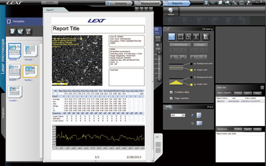
빠르고 이해하기 쉬운 보고서 작성
관찰 · 측정 결과의 신속한 보고서 작성도 레이저 현미경의 중요한 역할입니다. OLS4100은 측정 종료 후 한 번의 클릭으로 보고서 작성이 가능합니다. 개별 템플릿 사용자 지정이 자유 자재로 할 수있는 등 다양한 편집 기능도 갖추고 있습니다.
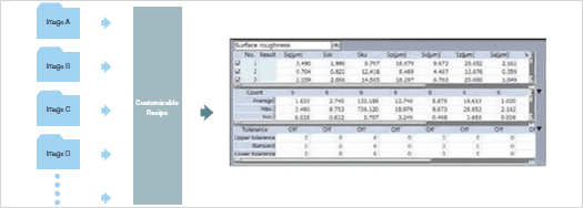
한번의 클릭으로 여러장의 이미지를 일괄 측정
상세 사용자 마법사 설정 기능은 긴 훈련의 필요 없이, 새로운 사용자도 빠르고 쉽게 작업을 할 수 있습니다.
OLS4100 Application
반도체/FPD
전자 부품 / MEMS
재료/금속 가공
반도체/FPD(Flat Panel Display)
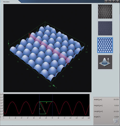
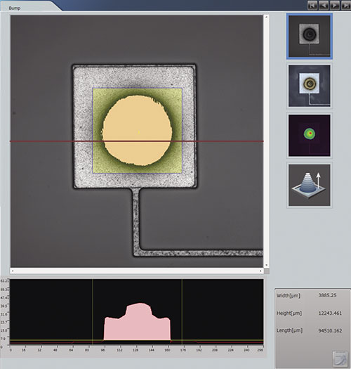
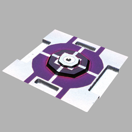
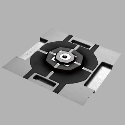
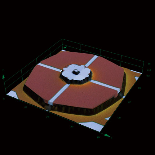
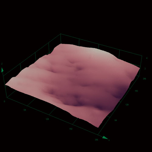
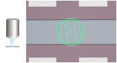
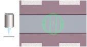
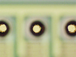
웨이퍼 범프 (대물 렌즈 100x/ 광학 줌 1.5x/스캐닝 영역 85 μm x 85 μm)
도광판 (대물 렌즈 50x/ 광학 줌 1x/스캐닝 영역 256 μm x 256 μm)
칩 패드 (대물 렌즈 50x/ 광학 줌 2x/스캐닝 영역 128 μm x 128 μm)
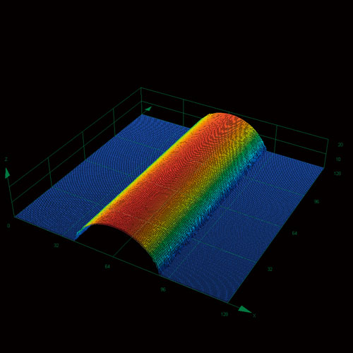
도광판 위의 레이저 점 (대물 렌즈 100x/ 광학 줌 1x/스캐닝 영역 128 μm x 128 μm)
전자 부품 / MEMS (미세 전자 기계 시스템)
포토 마스크 (대물 렌즈 20x/ 광학 줌 1x/스캐닝 영역 640 μm x 640 μm) 샘플 제공 : 코시부 정밀
마이크로 렌즈 (대물 렌즈 100x/ 광학 줌 1x/스캐닝 영역 128 μm x 128 μm)
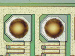
PCB 커넥터 (대물 렌즈 50x/ 광학 줌 1x/스캐닝 영역 256 μm x 256 μm)
MEMS (대물 렌즈 20x/ 광학 줌 1.3x/스캐닝 영역 483 μm x 483 μm)
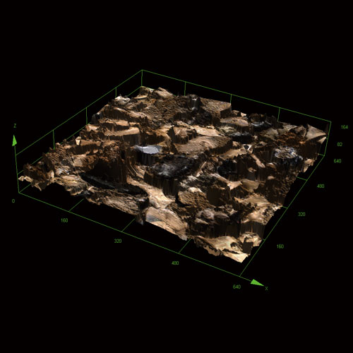
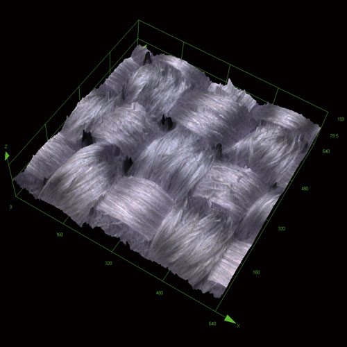
재료/금속 가공
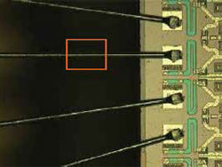
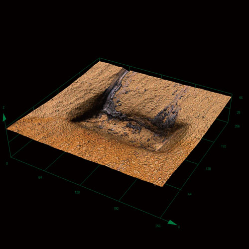
다이아몬드 전기 공구 (대물 렌즈 50x/ 광학 줌 1x/스캐닝 영역 256 μm x 256 μm)
탄소 (대물 렌즈 렌즈 100x/ 광학 줌 1x/스캐닝 영역 128 μm x 128 μm)
얇은 파이프 (대물 렌즈 렌즈 100x/ 광학 줌 1x/스캐닝 영역 128 μm x 128 μm)
접착 테이프 (대물 렌즈 50x/ 광학 줌 2x/스캐닝 영역 128 μm x 128 μm)
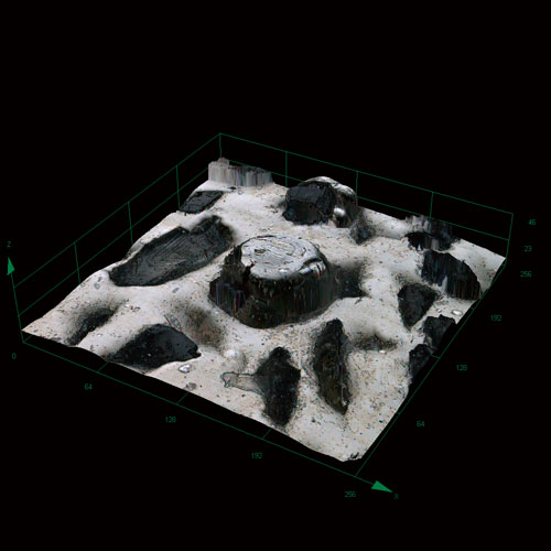
샌드 페이퍼 #400 (3D) (대물 렌즈 20x/ 광학 줌 1x/스캐닝 영역 640 μm x 640 μm)
대물 렌즈 20x/ 광학 줌 1x/스캐닝 영역 640 μm x 640 μm)
고밀도 구조(3D) (대물 렌즈 20x/ 광학 줌 1x/스캐닝 영역 640 μm x 640 μm)
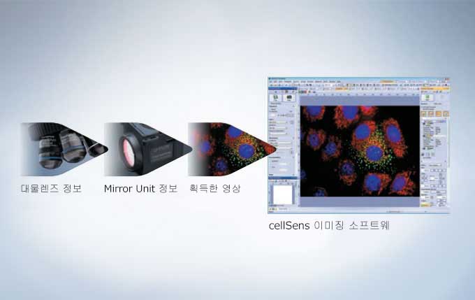
Olympus BX53 현미경은 cellSens 소프트웨어를 이용하여 조명 조절과 영상 획득이 가능하며 반자동 혹은 완전 자동화 옵션을 통해 실험에 대한 높은 유연성을 제공합니다. 그러므로 이러한 구성품들의 추가는 수준 높은 실험을 위해 뛰어난 가능성을 제공합니다.
보다 수준 높은 요구들을 충족시키기 위한 확장성
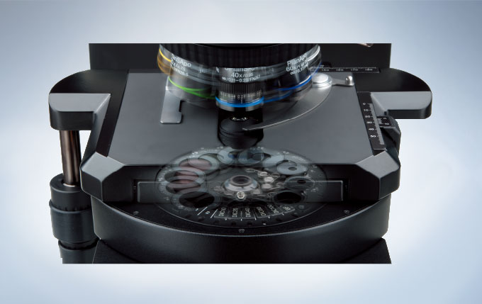
BX3 현미경 제품군은 연구 시장을 대상으로 폭 넓은 특징들과 우수한 광학 성능을 제공합니다. UIS2 광학계 시스템이 쉬운 조작의 전면부 제어기들을 채용한 탁월한 강도의 Y자 현미경 본체와 결합됨은, 다재다능함과 인체 공학적인 조작성을 제공합니다. 높은 공간 활용도의 BX53 현미경 본체는 다양한 작업과 함께 영상 처리를 통한 그 어떤 이미징 실험도 가능합니다. 그리고 디지털 이미징과 반자동 기능 그리고 형광(Fluorescence) 관찰의 완벽한 시작점이기도 합니다.
최적화된 명암 설정으로의 자동 전환
투과 조명 방식의 변경은 알맞은 맞는 대물렌즈 선택에 앞서 정확한 ND 필터, 편광판(Polarizer) / 분석판(Analyzer) 그리고 집광기 설정이 수반되어야 함을 의미합니다. 8구 범용 집광기는 모든 배율에서 사용할 수 있도록 상단 렌즈(Top lens) 까지도 조절합니다.. 이 집광기는 미분간섭(DIC) 프리즘, 위상차(Phase Contrast) 링슬릿과 편광판(Polarizer) 같은 광학 요소들의 쉬운 선택을 보증합니다. 조명과 대물렌즈를 잘 호환하기 위해, 광량 조리개는 자동으로 사용하고 있는 대물렌즈의 개구수로 설정됩니다.
설정 가능한 조절부 배열 형태
BX53은 중앙에 위치한 광량 조절 장치와 형광 조명 셔터를 어떤 손으로든 다룰 수 있게 하여 배치 시, 최대의 유연성을 발휘하도록 설계되었습니다. 또한, 미동나사는 핸들은 현미경 어느 측면에도 부착 가능합니다.
에너지-절약 스위치
동작감지기는 사용자의 부재를 파악하여 약 30분 뒤에 투과 조명 램프를 끕니다. 에너지 절약 스위치는 전력과 램프의 전력과 램프 수명을 아낄 수 있습니다.
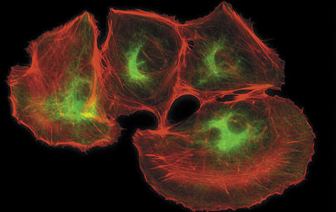
보다 향상된 감도의 형광 이미징
Olympus는 신호대잡음비(S/N ratio)를 극대화하고 형광(Fluorescence) 검출 능력을 개선하여, 밝은 시료의 색상과 어두운 배경으로 이루어진 형광(Fluorescence) 영상을 만듭니다. 균일한 조명과 검출 능력은 높은 투과율의 대물렌즈, 미러유닛과 fly-eye 렌즈 시스템의 결합으로 가능합니다.
Fly-eye 렌즈와 형광 조명장치들
시야 범위에 빛이 고르게 조사되도록 하는 것은 중요합니다. 형광 조명의 특성이 이를 더욱 어렵게 하지만, Olympus는 이를 해결하기 위해 fly-eye 렌즈 시스템을 결합한 새로운 형광 조명 기술을 개발하였습니다. 이러한 개선으로, 전 파장대의 균일한 조명뿐만 아니라 조명의 교정 또한 간단해졌습니다.
형광(Fluorescence) 관찰에서의 통합된 유연성
Olympus의 8구 형광 조명 장치는 매우 쉬운 교체가 가능한 미러 유닛들과 함께 다양한 형광(Fluorescence) 시료들을 위한 뛰어난 유연성을 제공합니다. 여러 색상의 형광(Fluorescence)을 보거나 FISH와 같은 실험 시의 미러 유닛 교체 필요성 감소를 통해 더욱 관찰이 빨라집니다.
개선된 코팅과 산란광 감소 기능의 형광 미러 유닛
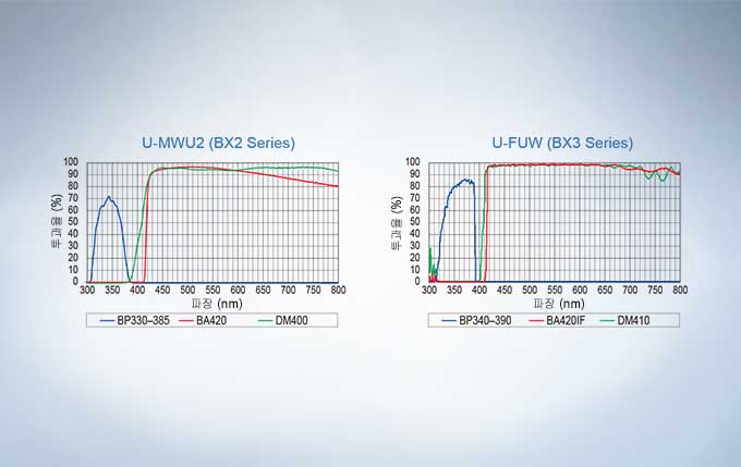
UIS2 형광(Fluorescence) 미러 유닛의 종류는 형광(Fluorescence) 영상 실험에 최적화 되었습니다. 고품질의 미러 큐브 코팅은 우수한 투과율과 수직에 가까운 파장 한계선을 제공함과 동시에 최상의 감도와 색분해능을 보장하기 위해, 내부 표면에서 발생하는 산란광을 99% 제거합니다. 미러 큐브들은 별도의 도구 없이 쉽고 빠르게 바꿀 수 있습니다.
자가형광(Autofluorescence) 저감을 통한 고 투과율
광학계의 품질은 광학 현미경의 핵심입니다. Olympus UIS2 광학계는 개발되어 정확도와 선명도의 기준을 선도합니다. 높은 NA값을 갖는 대물렌즈는 색수차를 보정하고 고해상도 영상 획득이 가능하여 약한 신호도 감지할 수 있습니다. Olympus는 선별된 유리 원료에 고급 UW 다중 코팅 기술을 적용하여, 대물렌즈의 자가형광(Fluorescence)을 감소하고 S/N ratio를 눈에 띄게 개선하였습니다. UW multi coating은 넓은 파장대에서의 고 투과율로 평탄한 영상을 보여 주며, 연구 용도의 여러 형광(Fluorescence) 색소의 최대 성능을 보장합니다.
배면반사(Back-Reflections) 저감을 위한 집광기 디자인
전동 전용 집광기는 배면반사(Back-Reflections)와 자가형광(Fluorescence)(Autofluorescence)을 감소하도록 설계되어, 형광(Fluorescence) 관측시 top lens를 swing-out할 수 있고, 자동으로 조리개(diaphragm)를 최소 축소하고, wheel을 두 위치 사이로 움직입니다.
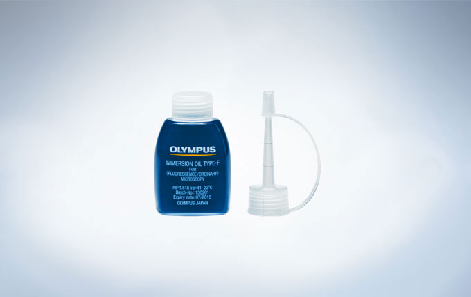
자가형광(Autofluorescence)이 감소된 이머전 오일
이 제품은 자가형광(Fluorescence)(Autofluorescence)을 저감하므로 형광(Fluorescence) 현미경용 이머전 오일에 적합하다. 감소된 노이즈(자가형광(Fluorescence), Autofluorescence)는 S/N ration를 향상시켜 우수한 형광(Fluorescence) 관측을 제공합니다. 자가형광(Fluorescence)(Autofluorescence)의 시간 변화가 적습니다. 노이즈가 생기기 쉬운 단일 분자 형광(Fluorescence)(single molecule fluorescence)의 정량 관측에 유용합니다. 결정화(Crystallization)에 대한 저항성은 장시간의 사용을 가능하게 합니다. 굴절 지표(Refraction Index)는 다른 Olympus 제품과 동일하며, 기존 현미경 시스템과 혼합하여 사용할 수 있습니다.
세계적 명성의 광학 성능
현미경 수준에서의 생물 표본은 명시야(Bright Field) 조명을 사용할 때 색상 편차와 같은 고유한 대비를 가지려 하지 않는 경향이 있습니다. 결과적으로 음영 대비를 만들기 위한 다양한 방식들이 개발되어왔습니다. 이들은 광학적 대조 방법과 시료를 통한 대조 방법, 이 2가지로 나눌 수 있습니다. 대조의 근원이 무엇이든지 간에 그 어떤 대조 방식에서도 Olympus의 BX3와 UIS2 광학 구성품들은 흠 잡을 데 없는 날카롭고 깨끗한 영상을 제공합니다.
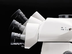
사용자에 맞춘 인체공학적 설계
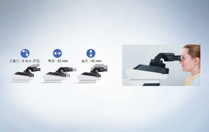
안정된 관측 자세는 사람마다 다르므로, 이 현미경은 각 사용자에 맞춰 mm 단위의 조절이 가능합니다. Olympus 경통 제품군은 경사각, 경통 연장 혹은 높이 조절과 같은 기능을 제공하여 완벽한 3차원적으로 유연한 조절이 가능합니다.
높은 조절성의 이안 경통
현미경 사용 시, 바르면서 편안한 자세를 취하는 것은 필수적이나, 전적으로 사용자에게 달려있는 부분입니다. 인체 공학적으로 완벽한 거리, 꺾임, 높이 조절이 가능한 경통은 경사각, 길이와 높이 조절이 가능, 3차원적으로 완벽하게 유연한 설정이 가능합니다. 결과적으로 현미경을 정확히 사용자에 맞게 조정할 수 있습니다.
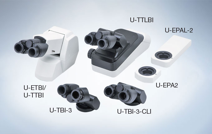
모든 요구를 만족하는 이안 경통
다양한 이안 경통 제품 군은 다양한 요구를 만족시킬 수 있습니다. 어떤 모델은 일반적인 도립상을 생성하는 반면 다른 모델은 표본과 동일한 방향으로 움직이는 정립상을 보여주어 표본 상에서 특정 부위를 보다 쉽게 찾을 수 있습니다.
회의와 이미징을 위한 직관적인 조절 방법
품질과 마찬가지로 모든 단계에서의 장비 호환성도 고려하여 설계되어야 합니다. 이를 목표로 Olympus는 연구용 정립 현미경 BX3 시리즈를 개발하였습니다. BX53은 최고로 다재다능한 영상 시스템을 대변합니다. 이는 사용 도중 진행되는 적응성을 포함, 수많은 광학 부품, 반자동과 소프트웨어 옵션들을 다채로운 선택을 제공합니다.
최적의 자세를 위한 꺾임형 삼안 경통
각 사용자는 고유한 자세 및 장비 배치에 대한 요구가 있으므로, 작업 환경의 개인화뿐만 아니라 현미경 조절을 통한 인체공학적 환경을 제공하는 것은 자세 혹은 반복되는 스트레스로 인한 부상이 없도록 하여 장기간의 현미경 사용이 가능하도록 합니다. 최대의 시스템 유연성 및 사용자 편이를 위해, 기울기 조절 가능한 인체 공학적 삼안 경통 튜브는 접안렌즈 높이 조절뿐만 아니라 눈동자 간 거리 조절이 가능하고, 광로 슬라이더는 경통 튜브 어느 쪽에도 부착이 가능하며, 사용자가 현미경 전체를 제어할 수 있도록 하고 자신의 자세에 맞춰 현미경을 조절하는 것을 보장합니다.
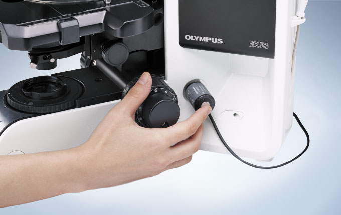
영상 획득용 핸드스위치
유선 노출 조절 장치는 현미경의 모든 면에 장착할 수 있습니다. 이는 모니터 방향으로 시선을 돌리고 마우스를 조작하는 과정 없이 간단히 단추를 누르는 것만으로 영상 획득을 가능하게 하여 사용자에게 더욱 효율적이고 인체공학적인 사용성을 제공합니다. 이는 더 효율적일 뿐만 아니라, 사용자를 더 좋은 환경을 제공하기 위해 인체공학적인 것입니다.
Coded Unit을 활용한 현미경 데이터 저장
BX3의 추가 구성품인 수동 노스피스(Nosepiece)와 형광필터 변경장치(Mirror turret) 모듈은 영상 획득 시, 자동으로 현미경의 배율과 설정 정보를 기록하고 공유할 수 있습니다. 이 판독(Readout) 기능은 Olympus의 cellSens 소프트웨어 패키지로 축척 오류 없는 영상을 기록할 수 있습니다.
늘어나는 요구에 최적화된 시스템
각 연구조사는 고유의 설정이 필요하기 때문에 연구용 현미경은 단순한 영상 획득을 위한 현미경을 넘어서야 합니다. 그 결과, 각 시스템은 높은 유연성 뿐만 아니라, 복잡 다양한 프로토콜과 프로세스들의 수행이 가능해야 합니다. Olympus BX3 시리즈 현미경은 호환성 높은 이미징 시스템과 함께 하드웨어와 소프트웨어를 사용자가 완벽히 조절할 수 있습니다.
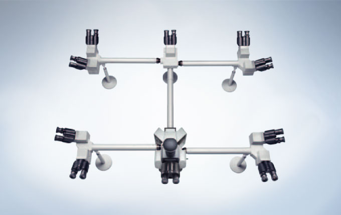
그룹 관측 시스템
인체 공학적인 이안 경통, 삼안 경통 뿐만 아니라 Olympus는 사용자에게 최적화된 실험실 토론을 위한 이인, 다중 관찰용 연결 장치를 제공합니다. 이 시스템은 모든 그룹이 각자의 접안렌즈로 동시에 표본을 관찰하면서 토론이 가능하므로 임상학적, 교육적 관찰에 매우 적합합니다. 폭 넓은 선택 가능성으로 2 ~ 10명 혹은 그 이상의 동시 관찰을 지원합니다.
광범위한 요구 충족을 위한 디지털 이미징
다재다능한 BX53 시스템은 어떤 어플리케이션에도 적용될 수 있는 자유로움을 제공하는 시스템 현미경 입니다. 최첨단 연구부터 컨퍼런스에 최적화된 독립형(Stand-alone) 모델까지 디지털 카메라 및 cellSens 이미징 소프트웨어의 모든 제품군은 고 S/N ratio의 형광(Fluorescence) 영상 획득을 보증합니다.
The STM7 microscopes offer excellent versatility and high performance three axis measurements of parts and electrical components, with sub-micron precision. Whether samples are small or large, simple or complex, or measurements are being taken by a novice or an expert, the Olympus STM7 range features measuring microscopes tailored to fit your needs.
Accurate Measurements through the Integration of an Optical Microscope and Advanced Measurement Capability
The STM7 was Designed with Emphasis on Ease of Use
Accessories that Widen the Range of Observation and Measurement
Automated Focusing System Provides Superior Repeatability
System for Achieving More Advanced Measurement
Accurate Measurements through the Integration of an Optical Microscope and Advanced Measurement Capability
Observation Performance Refined through Years of Microscope Development
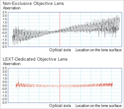
The STM7 series uses the same UIS2 infinity-corrected optical system used in state-of-the-art optical microscopes. As a result, observed images have high resolution and high contrast, with aberration thoroughly eliminated to help ensure highly accurate measurement in minute detail.
Measurement Reliability Enhanced with a Stage-Mounting Plate Crafted from Stone
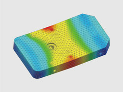
STM7-LF FEM analysis
To provide further assurance of measurement accuracy, the STM7 series uses a highly durable, vibration-resistant frame with a granite surface plate. As a result of this stability, measurements can be taken at sub-micron-levels while ensuring minimal error.
Continuing to Provide User-friendly, High-Precision, 3-axis Measurement as a Pioneer of Height Measurement
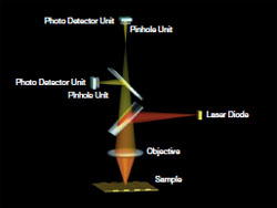
Reflective Active, Confocal Autofocus System Optical Path
As modern manufacturing technology becomes increasingly miniaturized and precise, highly accurate measurements are even more essential—not only along the horizontal XY axes, but also along the Z-axis. Olympus has responded to such needs by being the fi rst to realize an autofocus system for measuring microscopes by means of the reflective active, confocal method.
Dependable Quality Based On a Strict Traceability System
Measuring Microscopes Traceability System
The accuracy of Olympus’ measuring microscopes is controlled by a strict traceability system and Olympus even offers traceable calibration at the time of installation.
The STM7 was Designed with Emphasis on Ease of Use
Offering Stages to Fit the Sample Size at Hand
Common Problems
Short measurement stroke precludes the measurement of larger samples.
Sample rotation required to compensate for shorter Y than X-axis coverage during measurement is time inefficient. Until now, large stages have offered a sufficient measurement coverage on the X-axis, but only less coverage on the Y-axis.
Due to the narrow measurement range, it is impossible to line up large numbers of samples on the stage for measurement at once.
STM7 Solutions
Four types of stages are available, each with a unique square measurement stroke (choose from 50 mm x 50 mm, 100 mm x 100 mm, 200 mm x 200 mm, and 300 mm x 300 mm). From small to large size samples, there is a stage that fits the sample being measured.
A clutch system enables rapid switching between coarse and fine movements. Thanks to this switching function, the stage can also be moved rapidly along the X- and Y-axes, and freely across the XY plane.
The 300 mm square length stage enables the same measurement stroke to apply to both the X and Y-axes, which means it can be used to measure large samples, such as 300 mm wafers and printed circuit boards without changing their orientation.
Use the Same Microscope for Both Low- and High-Magnification Observations
Common Problems
Most conventional measuring microscopes only accept a measuring objective or metallurgical objective, and so are unable to meet the requirements for a wide variety of observations.
STM7 Solutions
The STM7 accepts both a metallurgical objective and a measuring objective by exchanging a revolving nosepiece with a measuring objective adapter. This means that the STM7 combines both metallurgical optics and measuring optics in one microscope. In this way, the STM7 series satisfi es a range of needs, no matter whether measuring a wide area or tiny region, measuring the size of differences betwe en levels, or assisting the user in deciding on the best observation method to choose.
Measuring Objectives
Because the measuring objectives have an extremely long working distance, they provide confidence when focusing on samples with large peaks and troughs while reducing worries of the objective coming into contact with the sample. Furthermore, their low-magnifi cation capability enables wide areas to be observed in a single view.
Metallurgical Objectives
Metallurgical objectives enable high-magnifi cation, highresolution observation capability comparable to that of optical microscopes. What’s more, these objectives can be used not only for brightfield, but also for darkfield and DIC observation.
> Click here for details about UIS2 objective lenses
Manual and Motorized Focusing Model Options
The STM7 Line Includes both Manual and Motorized Focus Options
Focus control is available with either manual or motorized operation. Choose the model that addresses your needs in terms of samples and measurement content, regardless of stage size—with all frames incorporating a linear scale for the Z-axis that enables 3-axis measurement.
Manual Z-axis Focus Models
Manual Z-axis focus models offer excellent cost performance—with familiar handle operation for rapid vertical movement that offers convenience for users who needs to measure samples with variety of heights.
Motorized Z-axis Focus Models
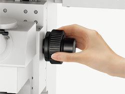
Operability is improved and handling fatigue is reduced for focus and height measurements when using the motorized focus unit. The coaxial knobs for coarse and fine movement offer a feeling similar to manual operation, while the models can also be equipped with an autofocus unit.
A Revolutionary Control Unit Refines Measuring Microscope Usability
Common Problems
Additional functions require additional operational units. Operators can’t always locate the corresponding unit quickly, which significantly reduces measurement efficiency.
Numerous operational units and their power supplies around the main unit occupy valuable working space.
STM7 Solutions
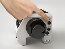
Controllers
With the STM7 series, a single controller makes it possible to perform virtually all measuring microscope operations, including use of readout reset, illumination control, focusing, and autofocus. For efficiency and convenience, the unit can be placed wherever you wish and operated easily with one hand.
Control Box
The power supply and transmission for each unit are combined in a single control box. This preserves maximal work space even when a range of optional functions, such as the focus navigator, are added.
Automatic Light Intensity Adjustment Greatly Improves the Efficiency of Observation and Measurement
Common Problems
Analog volume adjustment used by conventional measuring microscopes does not enable the quantitative assessment of light intensity, which can lead to variability in measured values as light intensity changes.
With conventional measuring microscopes, light intensity may need to be adjusted every time the objective is switched—making for an inefficient workflow.
STM7 Solutions
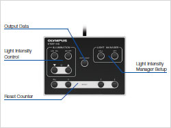
Close Control through a Quantitative Digital Display of Light Intensity Values
The STM7 series provides a quantitative digital display of light intensity—enabling observations to always be made under consistent illumination conditions.
Light Intensity Manager Eliminates the Need for Manual Adjustment
Light intensity manager can be used with the coded revolving nosepiece configuration. The coded revolving nosepiece automatically detects the switching of objectives. This allows the illumination method and light intensity to be registered for each objective, and adjusted automatically during measurements when the objective is switched. Now there is no need to manually adjust light intensity, which used to be required with every switch between magnifications.
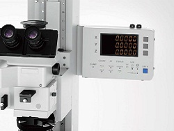
A Detachable Digital Read Out for Preferred Location Enables Swift, Convenient Checking of Measurement Results and Equipment Status
Common Problems
The need to check the operation status of equipment, such as illumination, or measured values on individual units makes overall operation cumbersome.
STM7 Solutions
Digital Indicator Enables the Current Operation Status to be Verified Visually
The indicator displays the device status and settings. The minimum X, Y, and Z-axis values can be switched between 0.1 μm and 1 μm, and the display units can be switched between mm, μm, inches and mil.
Detachable Digital Readout Allows for Individual Preference and Placement
Whether attached to the frame or a desk, the placement of the detachable digital readout is up to the individual user. While standing to take measurements, it can be placed on the side of the frame at almost the same height as the site of observation for an exceptional and easy view. When operating from a sitting position, such as observation or measurements on a monitor via a digital camera or when using the motorized Z-axis focusing model, simply place the digital readout and hand controller on the desk.
Automated Focusing System Provides Superior Repeatability
Achieve Faster, Simpler, More Accurate Height Measurement
Common Problems
When doing visual measurement, variations can arise in the height measurements between different operators. Furthermore, this measurement method is time-consuming and inefficient.
STM7 Solutions
Simple and Highly-Precise Focusing System with Superior Repeatability
The Olympus’ focus navigator delivers highly reproducible height measurement by projecting a pattern within the fi eld of view and identifying vertical deviations. Slight errors can occur in height measurements taken with normal visual observation, even when focus appears to be sharp. The focus navigator, however, enables measurements to be made simply by matching up the marks—thereby reducing operator subjectivity in measurement results.
Focus Navigator
Visual Height Measurement
Autofocus Advantage for Fast and Highly Accurate Height Measurement
Common Problems
During visual measurement, the results of height measurement can vary between different operators.
Manual height measurement requires the operator to repeatedly move the stage and adjust the focus with the handle, making measurement time-consuming and inefficient.
Focusing on minute objects, such as bonding wires, is difficult.
STM7 Solutions
Dedicated Autofocus Unit: Outstanding Reproducibility and Focusing Speed
The STM7 dedicated autofocus unit allows highly accurate height measurements to be made with minimal time, regardless of the level of operator experience. Use of the reflective active, confocal method provides a stable focal point independent of surface roughness or a slanting sample surface, while the small laser diameter enables the use of autofocus, even on minute objects, such as bonding wires.
One-shot Mode
Instantaneously takes autofocus from a roughly focused state to sharp focus located at the center of the field of view.
TRACK Mode
The featured TRACK Mode provides autofocus that tracks the peaks and troughs of the sample, even if the stage is moved, keeping the image continually in focus. This advancement greatly improves the efficiency of Z-axis measurements by enabling observations to be made without taking your hands off the X and Y handles.
Accessories that Widen the Range of Observation and Measurement
Coded Revolving Nosepiece
Combining a coded revolving nosepiece with a digital camera lets you display the objective magnification on-screen during observation and allows you to record that magnification. This convenient feature allows information on your sample and the objective’s magnification to be recorded at the same time when recording a sample.
MM6-EMO/ Erect Image Monocular Tube
Monocular tube for erect images. Can be used in combination with MM6-OCC10x (eyepiece with cross hairs).
STM7-FS/ Foot Switch
Enables hands-free transmission of data, allowing operators to complete measurement without taking hands off the X and Y handles.
SZ-LW61/ White LED Illumination Unit
This light-weight, space-saving design model provides a long operating life and low power consumption. The cost-effective LED illumination unit is also free from the flickering and brightness fluctuation.
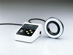
SZX2-ILR66+SZX-RHS/ LED Ring Illuminator+Manual Control Unit
SZX-RHS manual control unit enables independent illumination of four-segments of the SZX2-ILR66 reflected LED ring illuminator, which provides clear images with high color temperature. The optimal illumination can be selected from 13 patterns.
Rotatable Stage
Enables easy parallel alignment of sample.
System for Achieving More Advanced Measurement
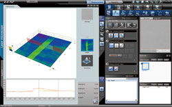
Measurement Support System
The ability to clearly and easily see the output display component of measuring microscopes is essential. That is why the new Olympus measuring software has been created, helping to deliver complex measurements with greater accuracy. The software also enables the use of digital cameras.
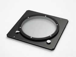
OLYMPUS SZX10은 Working Distance와 Field Size가 중요할 때 선택하시는 모델입니다. 렌즈의 디자인에 따라 왜곡현상없이 제품 혹은 샘플의 고유 색감을 그대로 관찰하실 수 있습니다.
안정성 및 반복성
인체공학적이고 빠른 사용
지능적인 디지털 영상
안정성 및 반복성
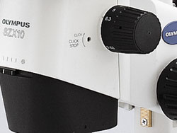
빠른 검사가 가능한 조절 손잡이
Zoom knob with click-stop
0.63배 에서 6.3배에 이르는 넓은 범위의 배율은 듀얼 렌즈 홀더를 통해 더 높은 배율도 볼 수 있으며, 별도의 렌즈 교체 없이 다양한 배율에서의 관찰이 가능합니다. 축을 이루는 빛 경로 설정의 장점은 카메라로 찍은 측정값이 현미경에서 샘플의 방향에 상관없이 모든 방향으로 안정적이고 정확하며 많은 영상 및 측정 작업들은 같은 배율에서 일관성있고 비교할 수 있는 정확한 결과값을 보장합니다. 11개의 click-stop 지점들은 중요한 기능들을 빠르고 쉽게 사용할 수 있도록 합니다.
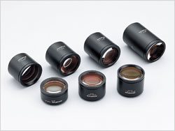
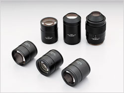
8개의 호환가능한 렌즈로부터의 뛰어난 성능
Lineup of Objective Lenses
DFPL2x/DPFL1.5x /DFPL 0.75x /DFPL 0.5x : DFPL 렌즈 시리즈는 뛰어난 색 재현성과 샘플의 모양을 정확하게 구현합니다.
DFPLAPO 1.25x /DFPLAP01x4 : 색 수차 및 구면 수차를 제거한 가장 높은 성능의 렌즈로 뛰어난 분해능과 contrast, 이미지 평면도와 최소한의 왜곡 등의 특징을 가집니다.
SZX-ACH1.25x /SZX-ACH1x : Long Working Distance와 고분해능을 갖춘 균형잡힌 렌즈입니다.
인체공학적이고 빠른 사용
인체공학적으로 편안한 사용 환경
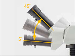
OLYMPUS는 인체공학적인 디자인이 사용자들에게 가장 중요한 요소라고 믿습니다. 따라서, 모든 동작은 사용하기 쉽고 눈이 편안한 접안렌즈, 낮은 위치의 스탠드 등으로 사용하는데 발생할 수 있는 스트레스를 최대한으로 줄였습니다. SZX2 실체현미경의 고급화된 OLYMPUS Comfort View 접안렌즈는 3D 이미지를 볼 수 있도록 넓은 범위를 한 번에 볼 수 있게하며 장시간 사용시에도 편안함을 가져다 줍니다. 또한 눈의 피로를 줄이며 실체 이미지를 더 쉽게 볼 수 있게 합니다. OLYMPUS는 튜브(헤드) 부분의 각도가 5~45로 조절할 수 있어 다양한 사용자들이 만족하며 사용할 수 있습니다.
자동기능
SZX10 전동 초점 장치는 화장 초점 영상(EFI)와 완벽하게 자동화된 디지털 문서를 만들 수 있습니다. 이러한 기능은 3D 이미지를 만들 수 있으며, 문서상의 이미지와 실제 눈으로 본 이미지의 격차를 줄여줍니다. 자동 검사를 용이하게하기 위해 OLYMPUS STREAM 이미징 소프트웨어 제품군은 간단한 2D 측정부터 복잡한 위상 분석에까지 다양한 환경에 사용하기 쉬운 모든 도구들을 제공합니다.
다양한 종류의 스탠드와 조명 장치
Low-profile LED Illumination Base
OLYMPUS SZX2는 OLYMPUS LED 기반의 특별한 시스템을 갖추고 있어 투과 조명의 미세한 조정으로 밝기와 영상을 컨트롤 하실 수 있습니다. 울트라 슬림의 40mm Base와 함께 SZX2는 가장 인체공학적인 높이의 스테이지를 지원합니다. 게다가 명시야, 암시야, Oblique 조명의 선택을 회전식 스위치를 통해 쉽게 할 수 있습니다. 긴 수명의 LED는 샘플이나 스테이지에 열을 전달하지 않으며 Material 실체현미경으로써의 유연성을 강화하였습니다.
다양한 종류의 반사형 조명 장치
링 라이트 방식의 조명이나 동축 조명등의 다양한 반사형 조명유닛들의 사용이 가능합니다. 사용자의 어플리케이션에 맞는 최적의 조명 시스템을 선택하실 수 있습니다.
지능적인 디지털 영상
디지털 영상
SZX16과 함께 당신은 샘플의 실시간 디지털 영상을 보여주거나 문서를 제작하고 싶어질 것입니다. OLYMPUS 쿨링 방식의 DP를 같이 사용하여 매우 유용한 이미징 스테이션을 구착할 수 있습니다. 이 것은 빠른 속도의 라이비 영상 관찰 뿐만 아니라 고해상도(1730)만화소까지)의 문서 제작도 가능합니다. 발전된 인터페이스는 샘플이 움직이는 동안에도 색재현성이 뛰어나고 색변화가 자유롭습니다.
OLYMPUS의 정교한 현미경 디지털 카메라 및 이미지 분석 소프트웨어 패키지는 관찰, 리포트 생성, 데이터베이스, 보관 및 SZX16 전동 줌 초점 제어를 포함한 다수를 작업을 지원합니다. OLYMPUS가 디지털 카메라 및 분석 / 측정 소프트웨어의 전체 라인을 제공하며, 고객님의 OLYMPUS 담당자는 사용자의 요구 사항에 맞춰 충족시켜드릴 수 있도록 도와 드릴 것입니다.
Work Comfortably and Productively
Natural Posture, Reduced Stress, and Increased Productivity
The ergonomic long tilting trinocular provides an optimized work position by bringing the microscope closer to the user while the extendable eyepoint adjuster provides flexibility for users of different heights. The SZX series’ ergonomic instruments reduce stress during observation by providing the most comfortable position for each user, increasing work efficiency.
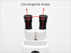
Convergence Angle in Tube Relieves Eyestrain
Olympus research established a correlation between stereo microscope optical systems and eyestrain. Certain convergence angles between the left and right optical paths can cause discomfort. The SZX2 series is designed with a convergence angle that completely compensates for each optical path. This solution effectively eliminates eyestrain, even during prolonged observation.
Ergonomic Zoom and Focus Knobs for Fatigue-free Use
The position of the zoom knob, size and position of the coarse/fine focusing knob, and the fine focus stroke are designed for easy operation. The enhanced fine focus stroke results in easy and precise focusing.
Slim Design Illumination Stand for Easy Access to Samples
Illumination stands are designed to be easy to use and fatigue-free. The slim LED transmitted light illumination stand, at approximately 40 mm in height, features easily adjustable fingertip illumination control and provides easy access to samples.
Specification
Research Stereomicroscope System SZX10 Specifications
Focusing Unit/Coarse Fine Focusing Unit/Heavy-duty Coarse Fine Focusing Unit/Motorized Focusing Unit
Objective Lens
Magnification
Type
N.A.
W.D. (mm)
Total Magnification *2 *3
Field Diameter of View (mm)*2 *3
0.5x
Plan Achromat
0.05
171
3.2x-31.5x
Ø69.8-Ø7
0.75x
Plan Achromat
0.075
116
4.7x-47.3x
Ø46.6-Ø4.7
1x
Plan Apochromat
0.1
81
6.3x-63x
Ø34.9-Ø3.5
Achromat
0.1
90
6.3x-63x
Ø34.9-Ø3.5
1.25x
Plan Apochromat
0.125
60
7.9x-78.9x
Ø27.9-Ø2.8
Achromat
0.125
68
7.9x-78.9x
Ø27.9-Ø2.8
1.5x
Plan Achromat
0.15
45.5
9.5x-94.5x
Ø23.3-Ø2.3
2x
Plan Achromat
0.2
33.5
12.6x-126x
Ø17.5-Ø1.7
Dimensions (W x D x H)
285 mm x 335 mm x 400 mm
Weight
7 kg (in Standard Configularion)
Remark
*1 total magnification range possible by combining an objective lens and eyepiece *2 in the case of using eyepiece 10x *3 SZX2-LTTR: intermediate magnification is 1.25X, SZX2-ILLC16/10: intermediate magnification is 1.5X
OLYMPUS SZX16은 900 line pair/mm 의 높은 분해능을 가지고 있어 고사양의 성능을 필요로 하는 어플리케이션에 사용될 수 있도록 제작되었습니다. 0.7x – 11.5x 에 이르는 줌 배율과 듀얼터렛을 사용하여 2개의 대물렌즈를 사용하실 수 있습니다.
광학의 우수성
인체공학적이고 빠른 사용
지능적인 디지털 영상
광학의 우수성
넓은 줌 범위
0.7배에서 11.5배에 이르는 넓은 줌 범위를 갖고 있어 추가적인 대물렌즈의 변경없이 넓은 시야에서 부터 자세한 부분까지 관찰이 가능합니다.
다양한 종류의 대물렌즈
Lineup of Objective Lenses
SDF 시리즈의 대물렌즈와 함께 비점수차를 제거하여 높은 분해능과 contrast를 가진 이미지를 관찰 할 수 있습니다. 샘플의 요구사항에 맞게 6가지 종류의 렌즈를 통해 가장 적합한 대물렌즈를 선택하여 사용할 수 있습니다. parfocal 렌즈의 가장 넓은 범위 (0.5x, 1.0x, 1.6x, 2.0x )와 함께 큰 줌 비율은 SZX16가 macro-view부터 micro-view까지 관찰할 수 있게 합니다. 놀라운 분해능과 배율의 선택은 작업을 더 효율적으로 하고 샘플로부터 더 정확한 정보를 얻을 수 있습니다.
인체공학적이고 빠른 사용
자동기능
SZX16 전동 초점 장치는 화장 초점 영상(EFI)와 완벽하게 자동화된 디지털 문서를 만들 수 있습니다. 이러한 기능은 3D 이미지를 만들 수 있으며, 문서상의 이미지와 실제 눈으로 본 이미지의 격차를 줄여줍니다. 자동 검사를 용이하게하기 위해 OLYMPUS STREAM 이미징 소프트웨어 제품군은 간단한 2D 측정부터 복잡한 위상 분석에까지 다양한 환경에 사용하기 쉬운 모든 도구들을 제공합니다.
인체공학
OLYMPUS는 인체공학적인 디자인이 사용자들에게 가장 중요한 요소라고 믿습니다. 따라서, 모든 동작은 사용하기 쉽고 눈이 편안한 접안렌즈, 낮은 위치의 스탠드 등으로 사용하는데 발생할 수 있는 스트레스를 최대한으로 줄였습니다. SZX2 실체현미경의 고급화된 OLYMPUS Comfort View 접안렌즈는 3D 이미지를 볼 수 있도록 넓은 범위를 한 번에 볼 수 있게하며 장시간 사용시에도 편안함을 가져다 줍니다. 또한 눈의 피로를 줄이며 실체 이미지를 더 쉽게 볼 수 있게 합니다. OLYMPUS는 튜브(헤드) 부분의 각도가 5~45로 조절할 수 있어 다양한 사용자들이 만족하며 사용할 수 있습니다.
Viewing tube (head) of adjustable angle
Adjustable observation tube(head)
다양한 종류의 스탠드와 조명 장치
OLYMPUS SZX2는 OLYMPUS LED 기반의 특별한 시스템을 갖추고 있어 투과 조명의 미세한 조정으로 밝기와 영상을 컨트롤 하실 수 있습니다.울트라 슬림의 40mm Base와 함께 SZX2는 가장 인체공학적인 높이의 스테이지를 지원합니다. 게다가 명시야,암시야, Oblique 조명의 선택을 회전식 스위치를 통해 쉽게 할 수 있습니다. 긴 수명의 LED는 샘플이나 스테이지에 열을 전달하지 않으며 Material 실체현미경으로써의유연성을 강화하였습니다.
Low-profile LED Illumination Base
다양한 종류의 반사형 조명 장치
링 라이트 방식의 조명이나 동축 조명등의 다양한 반사형 조명유닛들의 사용이 가능합니다. 사용자의 어플리케이션에 맞는 최적의 조명 시스템을 선택하실 수 있습니다.
Work Comfortably and Productively
Natural Posture, Reduced Stress, and Increased Productivity
The ergonomic long tilting trinocular provides an optimized work position by bringing the microscope closer to the user while the extendable eyepoint adjuster provides flexibility for users of different heights. The SZX series’ ergonomic instruments reduce stress during observation by providing the most comfortable position for each user, increasing work efficiency.
Convergence Angle in Tube Relieves Eyestrain
Olympus research established a correlation between stereo microscope optical systems and eyestrain. Certain convergence angles between the left and right optical paths can cause discomfort. The SZX2 series is designed with a convergence angle that completely compensates for each optical path. This solution effectively eliminates eyestrain, even during prolonged observation.
Ergonomic Zoom and Focus Knobs for Fatigue-free Use
The position of the zoom knob, size and position of the coarse/fine focusing knob, and the fine focus stroke are designed for easy operation. The enhanced fine focus stroke results in easy and precise focusing.
Slim Design Illumination Stand for Easy Access to Samples
Illumination stands are designed to be easy to use and fatigue-free. The slim LED transmitted light illumination stand, at approximately 40 mm in height, features easily adjustable fingertip illumination control and provides easy access to samples.
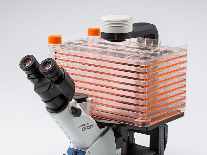
향상된 이미지 품질과 인체공학 설계로, Olympus CKX53은 라이브 셀 관찰, 세포 샘플링 및 처리, 이미지 캡처, 그리고 형광 관찰을 포함한 다양한 세포 배양 샘플에 뛰어난 성능과 효율적인 관찰 흐름을 제공합니다.
라이브 셀 관찰
통합 위상차( iPC : Integrated Phase Contrast ) 현미경
CKX53 iPC 시스템을 이용하여 대물렌즈 배율( 4x, 10x, 20x , 40x ) 변경시, 콘덴서 측의 위상차 링슬릿의 연동 변경이 필요 없게 되어 효율적인 관찰 작업이 가능하고, 또한 위상차 설정이 틀어짐에 대한 수시 조정이 필요없어 언제나 선명한 샘플 관찰이 가능합니다.
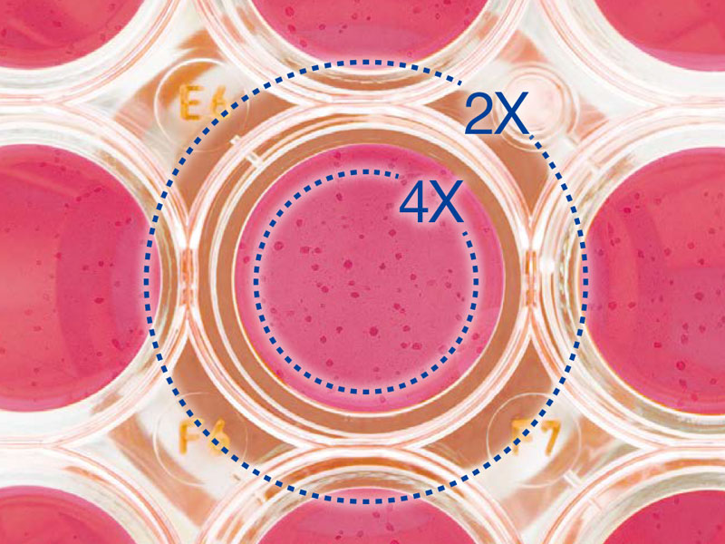
2X 배율, FN 22의 대물렌즈로 선명하고 넓은 시야
PLN2X 대물렌즈를 위한 링 슬릿, CKX3-SLPAS는 직경 11 ㎜ , 시야수 22 ㎜ 를 갖습니다.
2X 대물렌즈는 다른 대물렌즈 보다 확연히 높은 contrast를 제공하여, 투명한 샘플도 명확하게 식별할 수 있습니다. 예를 들어, 96-웰 마이크로 플레이트 관찰시, 넓은 시야로 인하여 스테이지를 움직이지 않고 웰의 모든 세포를 관찰 할 수 있습니다.
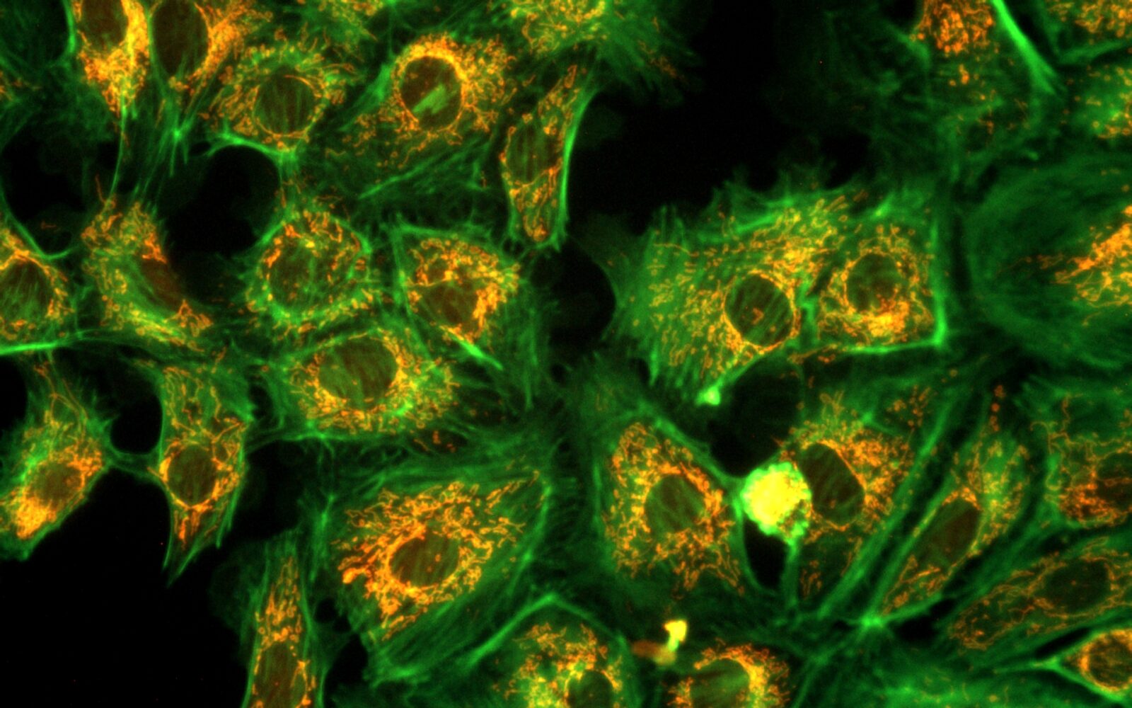
IVC (Inversion Contrast) 기술을 사용한 3D 셀 관찰
새로 개발된 IVC 기술로, 위상차보다 시야 심도는 좁아지며, 개체의 모양이나 투명도와 관계없이 삼차원 이미지를 선명하게 합니다. 또한, IVC 관찰은 후광 효과나, 방향성 있는 그림자를 배제하여, 개체의 선명한 관찰을 가능하게 합니다. * 10X 대물렌즈 (PLCN10X, CACHN10XIPC)는 새로운 IVC 관찰에 사용할 수 있습니다.
Glass Heater for microscope
TPi-CKX53X ( Thermo Glass Plate )
Microscope:Olympus CKX53 series
Applicable stage: XY mechanical stage CKX3-MVR
Setting range: ambient ~ 60℃
Plate dimension: W190 x D138㎜
Heating area: W174 × D127㎜
Glass thickness: 0.5 ㎜
형광 관찰 (Fluorescence Microscopy)
다양한 형광 시약과 선명한 시야
100 W 수은 램프 (U-LH100HG), 130 W 고압 수은 램프 (U-HGLGPS), 그리고 타사(3rd Party) LEDs*와 같은 여러 통합 광원을 이용하여 형광 이미지를 선명하게 관찰할 수 있습니다. 일반 연구용 형광현미경 IX3 및 BX3 에서 사용하는 미러 유닛을 동일하게 사용 할 수 있습니다.
3개의 형광미러 유닛을 장착할 수 있으나, Bright Field 관찰과 위상차 관찰에 영향을 줄 수 있으니 유닛 선택시 고려할 필요가 있습니다.
Image taken by AcquCAM 23GR2 with LUCPlanFLN40x Ph2, 1x Adapter, CKX53
밝은 조건에서 높은 Contrast
“Umbra Shield”는 특히 CKX53을 사용한 형광 관찰을 위해 설계되었습니다. 차단막은 실내 광원을 효과적으로 차단하여 형광의 대비를 향상하여 밝은 실험실 조건에서도 선명한 형광 관찰이 가능합니다. 위상차를 사용하는 경우, Umbra 차단막을 들어 올려 표본에 빛을 통과시킬 수 있습니다.
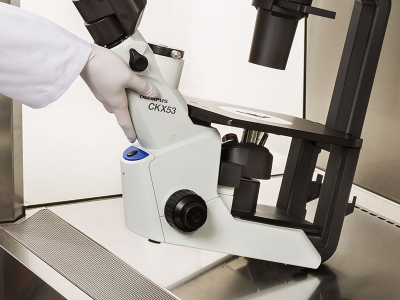
CKX53 현미경은 UV 차단 코팅 덕분에 UV 살균 공정 중에 그대로 둘 수 있습니다. 이 시스템은 약 7kg (15.4lb)으로 이전 모델보다 가볍고 설치 공간이 더 작기 때문에 실험실 공간을 덜 차지합니다. 또한, 한 손으로 현미경을 움직일 수 있으며 관찰 경통의 목 부분을 이용하여 쉽게 운반 할 수 있습니다.
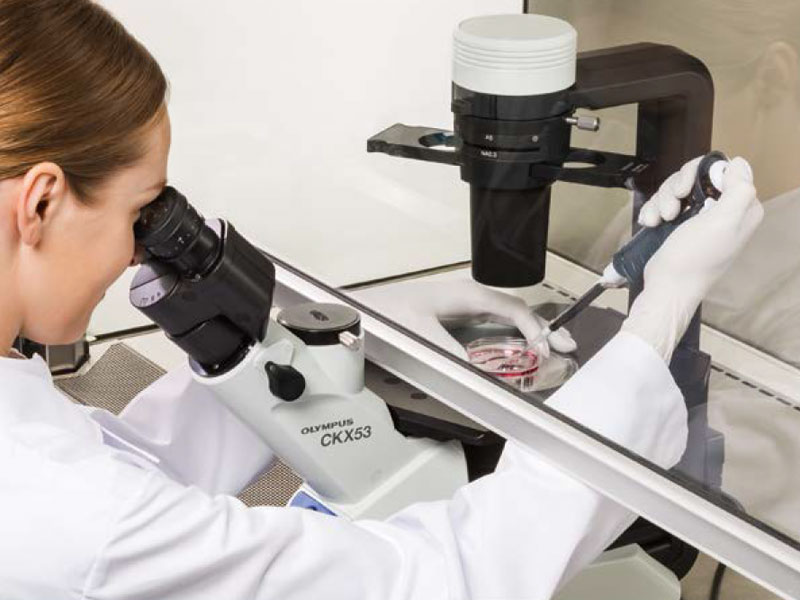
멸균 벤치 환경에서 간편한 세포 샘플링
CKX53의 접안렌즈와 광축/포커스 노브 사이의 거리가 짧으므로 작업자의 손의 위치를 자연스럽게 잡을 수 있어서 초점 및 셀 샘플링이 용이합니다.
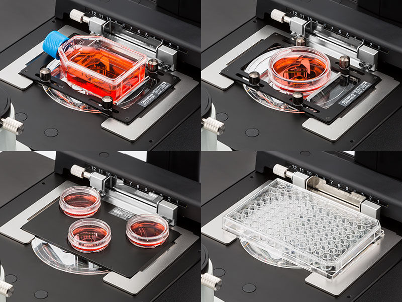
다양한 세포 배양 용기를 사용할 수 있습니다.
CKX53의 공용 홀더로, 디쉬, 마이크로플레이트, 플라스크를 포함한 다양한 용기에서 배양된 세포를 확인하기 쉽습니다. 옵션 홀더가 부착되면, 최대 세 개의 35㎜ 배양 용기를 스테이지에 장착할 수 있습니다. 또한, 다양한 마이크로플레이트를 별도의 홀더 없이 다룰 수 있습니다.
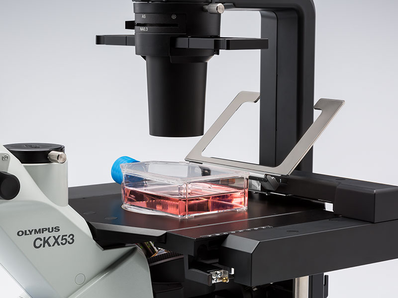
다층 조직 플라스크(Multi-Layer Tissue Flask)를 위한 종합적인 관찰
CKX53의 폭과 탈착 가능한 콘덴서로 다층 조직 플라스크와 같은, 최대 190 ㎜ 높이의 배양 용기도 볼 수 있습니다. PLCN4X 대물렌즈의 우수한 초점 심도로 다층 조직 플라스크 내 바닥 두 개의 층의 세포를 빠르고 편하게 관찰할 수 있습니다.
다양한 용기를 사용하여 관찰 유연성 증대
홀더 암을 들어 올려서 수동으로 세포 배양 용기를 배치할 수 있습니다. 또한, 스테이지는 좌우로 최대 70 ㎜ 까지 확장할 수 있습니다.


