Patent registration: Observation method and observation device
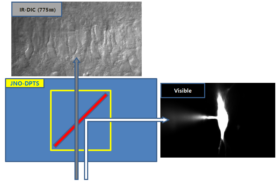
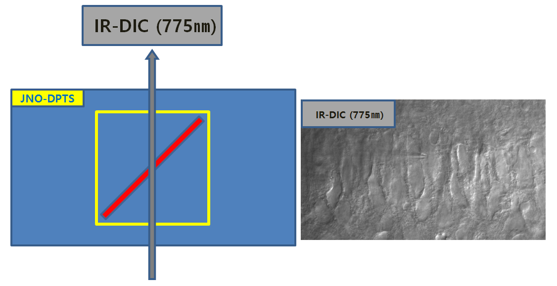
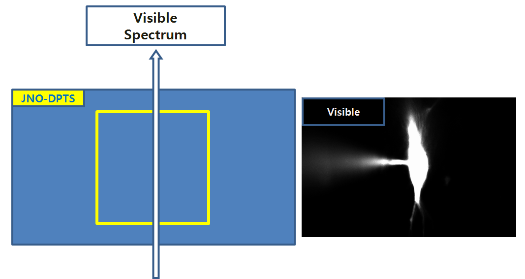
Simultaneous observation ( Bright Field and Fluorescence microscopy)
Fluorescene Microscopy (형광 현미경 검경법)과 Bright Field Microscopy (명시야 현미경 검경법)의 동시관찰을 위한 관찰 방법 및 관찰 장치
March 09, 2021
Patent registration: sample fixing device and method of placing sample
Patent applied to section observation microscope (JNO-FM-BX53-SET)
Sample holding device and method of placing sample Sample fixing device for observing the cross section of the flexible packaging film and method of placing the sample (Patent)
June 12, 2014
Patent registration: Method of measuring the height of a sample using a microscope
Patent: Height measurement method of sample by microscope
현미경을 이용한 샘플의 높이측정방법
June 5th ~ June 15th, 2000 ( OLYMPUS Japan in Tokyo)
Basic Knowledge of Microscope and Repair Training Course
September 4, 2003 ( OLYMPUS Japan in Tokyo)
Measuring Microscope Calibration License
Main Model: STM6
License ID Number: M029
2004/11/302003 /10/01
September 4, 2003 ( OLYMPUS Japan in Tokyo)
Biological Confocal Microscope Setup & Service Training
Main Model: FluoView300 & FluoView500
October 3, 2003 ( OLYMPUS Japan in Tokyo)
Inverted Microscope & Research Stereo Microscope Service Training
Main Model: IX71 / SZX9
August 17th ~ August 20th, 2004 ( OLYMPUS Japan in Tokyo)
Confocal Microscope Setup & Service Training
Main Model: FV1000
October 11 ~ October 13, 2004 ( OLYMPUS Japan in Tokyo)
Confocal Microscope Setup & Service Training
Main Model: FV1000 M-COMB(Multi Combiner)
June 20, 2005 ~ June 23, 2005 ( OLYMPUS Japan in Tokyo)
Research System Microscope Service Training
Main Model: AX70
August 1st-August 2nd, 2005 ( OLYMPUS Japan in Tokyo)
Polarizing Microscope Instruction Training
Main Model: BX51P
December 19, 2005 ( OLYMPUS Japan in Tokyo)
ZDC Training for Maintenance & Basic Knowledge of Confocal Microscope
December 20 ~ 22, 2005 ( OLYMPUS Japan in Tokyo)
Brushup Course to bring out the power of a microscope (GA Academy)
August 2nd ~ August 4th, 2006 ( OLYMPUS Japan in Tokyo)
Confocal Laser Scanning Microscope for Industrial Market
D class License(for ols3000)
Main Model: OLS 3000(Lext)
October 11 ~ October 15, 2006 ( OLYMPUS Japan in Tokyo)
Confocal Laser Scanning Microscope Training for Bio Maket
Maintenance Service Training
Main Mode : M-COMB(Multi Combiner for FV1000)
December 9, 2006
IX Repair training
May 07 ~ May 11, 2007 ( OLYMPUS Japan in Tokyo)
Modify Training for Zero-Drift Compensation Unit of IX81
IX2-Cusominsing Training (Optic Port Modify)
Main Model: IX71 & IX81-ZDC
September 3rd ~ September 08th, 2007 ( OLYMPUS Japan in Tokyo)
Leicense D class Trainging
Main Model: OLYMPUS Bio Confocal Microscope FV1000
April 08 ~ April 10, 2008 ( OLYMPUS Japan in INA_Nagano )
IV100 Setup Training
(in vivo fluorescence molecular imaging systems)
July 22 ~ July 23, 2008 ( Narishige Group in Tokyo, Japan )
Narishige Maintenance Training
July 24th ~ July 28th, 2008 ( OLYMPUS Japan in Tokyo)
Research Inverted Microscope maintenance Training
Main Model: IX81
May 18 ~ May 22, 2009 ( OLYMPUS Japan in Tokyo)
OLYMPUS Bio Confocal Microscope License Training
License Grade: FV1000 C class
June 23 ~ June 26, 2009 ( OLYMPUS Japan in Tokyo)
Confocal Laser Scanning Microscope License Training
License Grade: FV10i C class
October 19 ~ October 23, 2009 ( OLYMPUS Japan in Tokyo )
Multi Photon Laser Scanning MICROSCOPE Setup Training
Main Model: MPE C class
August 3rd ~ August 8th, 2010 ( OLYMPUS Japan in Tokyo )
Modify Training for Zero-Drift Compensation Unit of IX81
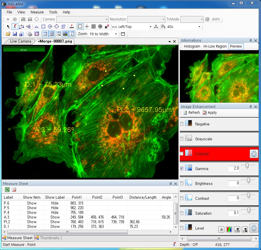
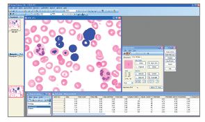
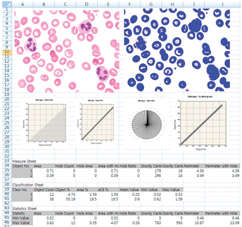
ARM is image analysis software for JNOPTIC AcquCAM cameras. This S/W is interchangeable with all WDM cameras regardless of camera brand and model and even more the most strength point is simple and easy use of length, area, angle, etc.
2. Simple measurement tools
Measurement Tools: Count, Distance, Angle, Area, etc.
Available measurement on LIVE image and saved image mode
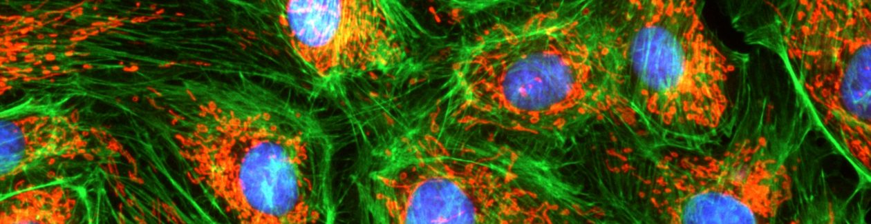
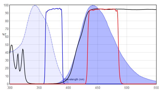
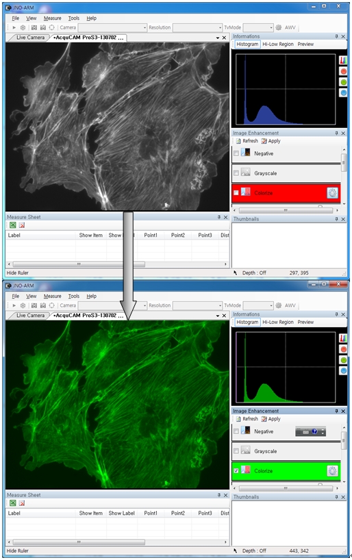
3-1. Specialization for observation of fluorescence images
Improvement Function about Fluorescence Microscopy Image. (Used function: Auto Level, Low level, Pseudo Color, Merge Image)
Bottom on the left is merged image without improvement of image. And the bottom on the right is merged image after improvement of image. These images are shot by JNOPTIC AcquCAM Pro/G3 camera
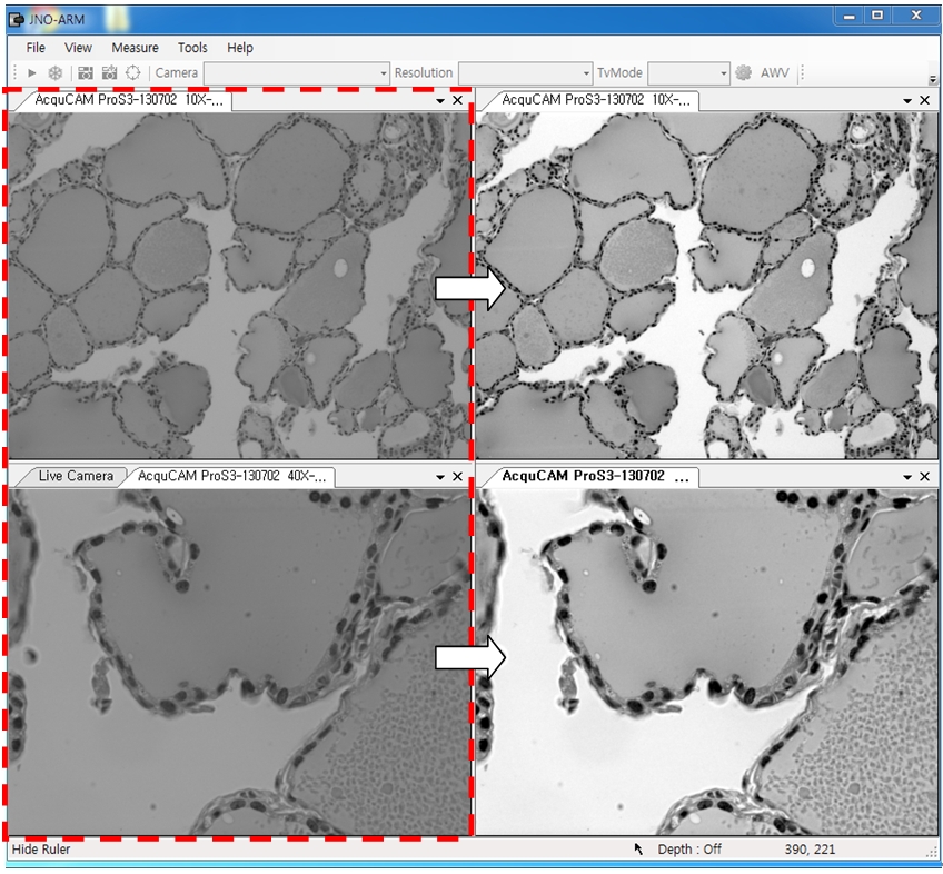
3-2-1 Effect of high-contrast image with simple operation (Monochrome)
Improvement Function about Microscopy Image Monochrome. (Used function: Auto Level)Effective improvement of image with simple operation. This image is shot by JNOPTIC Pro/S3 camera
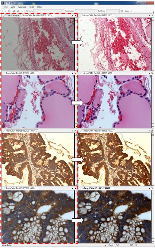
3-2-2 Effect of high-contrast image with simple operation (Color)
Improvement Function about Microscopy Image. (Used function: Auto Level)Effective improvement of image with simple operation. These images are shot by JNOPTIC AcquCAM Pro/G3 camera
3-3-1 Live Pseudo Color (for color camera)
When you get fluorescence images using color camera, if the unwanted channel was shown because of cross talk, you could remove this channel to take advantage of function of Live Pseudo.
Improvement Function about Fluorescence Microscopy Live Camera Image. (Used function: Live Pseudo Color)
The extraction of wanted color information from color image (Remove BR on RGB source) These images are shot by JNOPTIC AcquCAM Pro/G3 camera
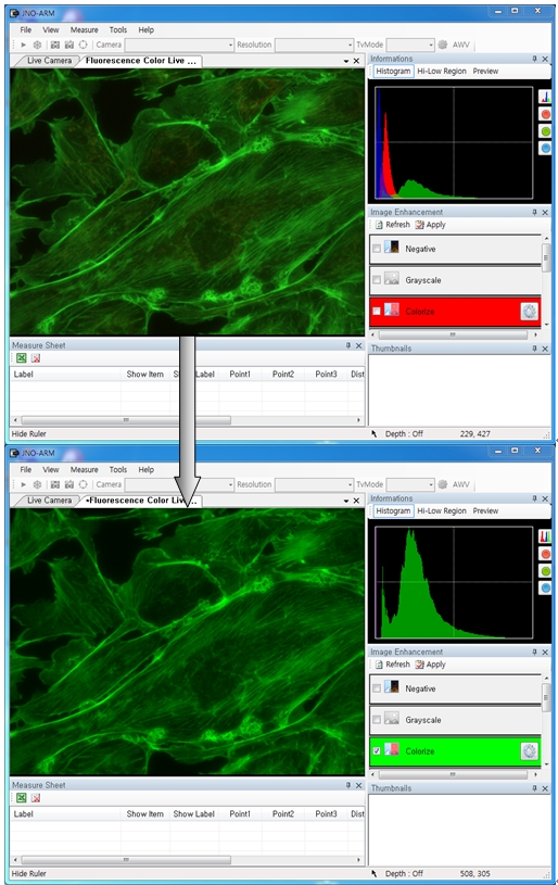
3-3-2 Live Pseudo Color (for Monochrome camera)
When you get fluorescence images using monochrome camera, you can raise efficiency of acquisition for fluorescence images to add similar false color to the color to be shown on the eyepiece in real time.
Improvement Function about Fluorescence Microscopy Live Monochrome Camera Image. (Used function: Live Pseudo Color)
Improvement of usage environment for monochrome camera to add false color on the image. These images are shot by JNOPTIC AcquCAM Pro/S3 camera
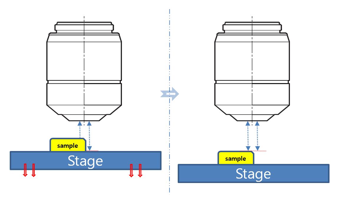
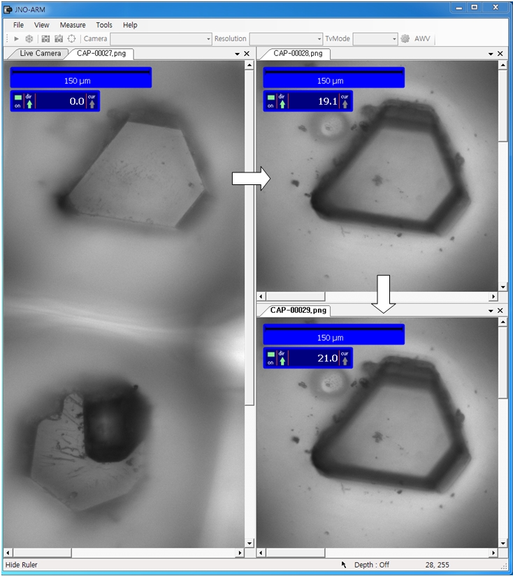
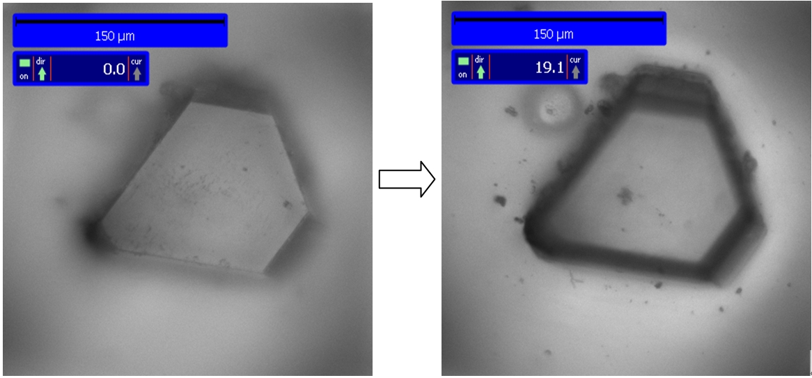
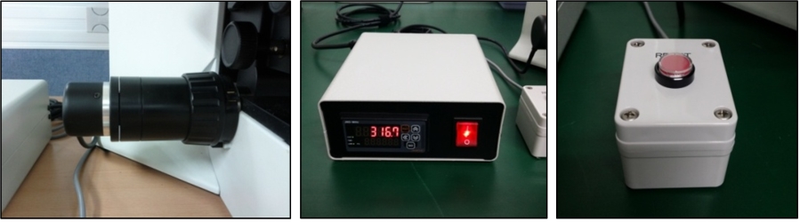
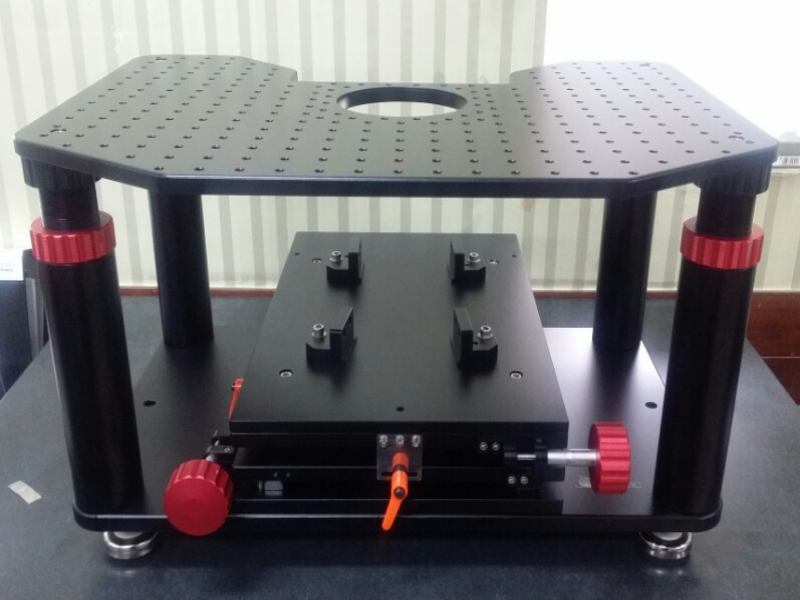
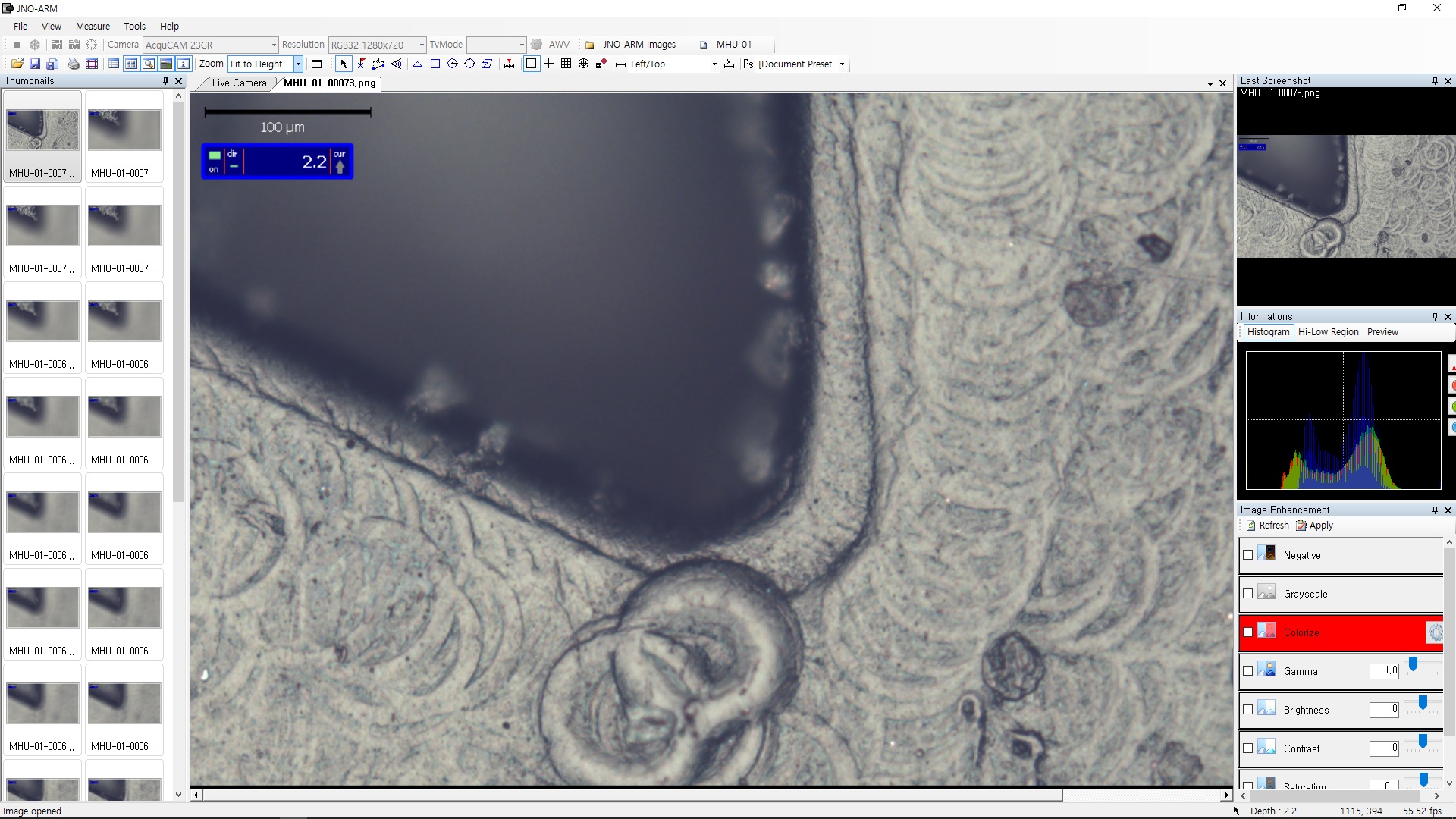
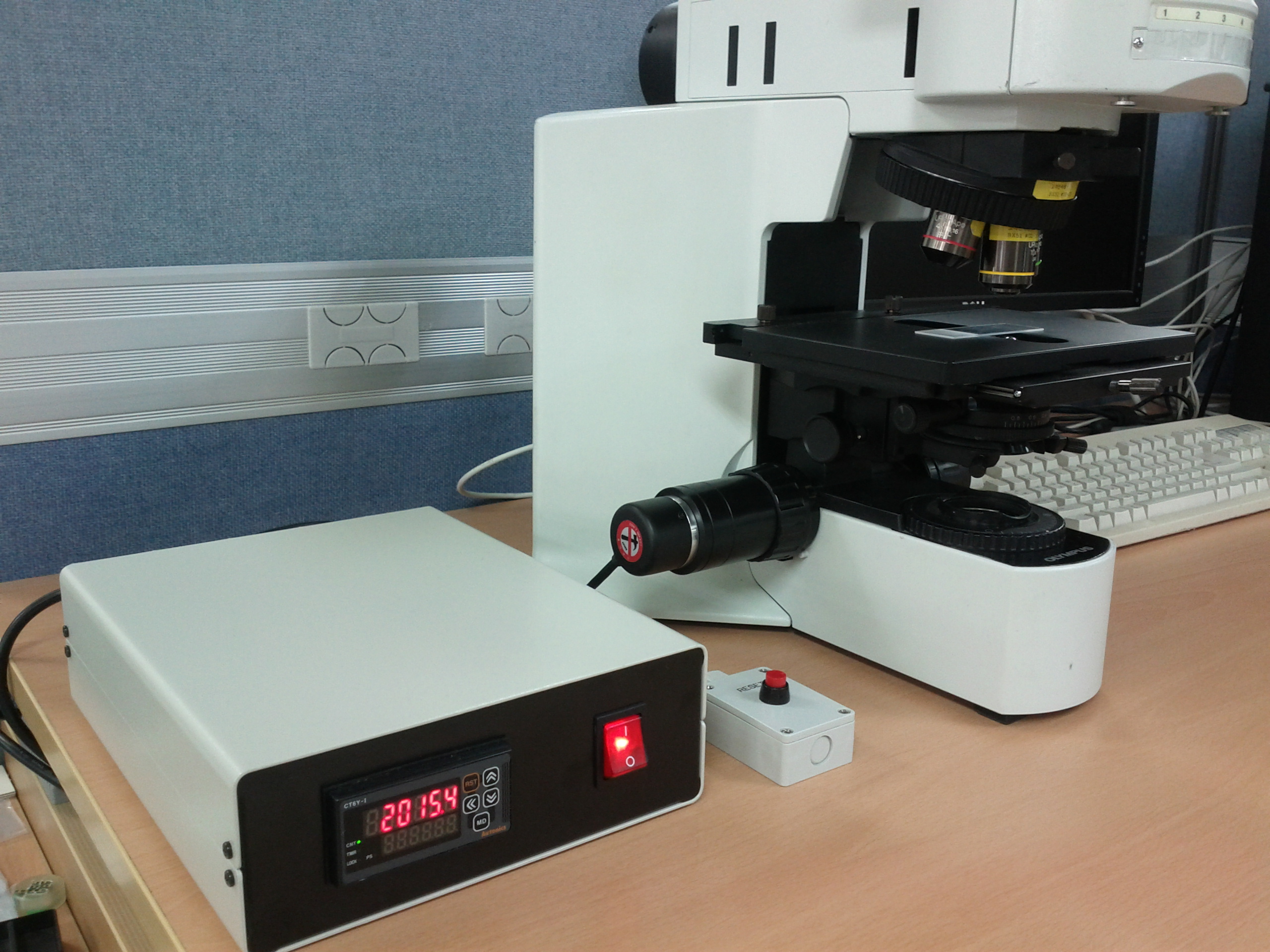
4. JNO-MHU(Option Unit)
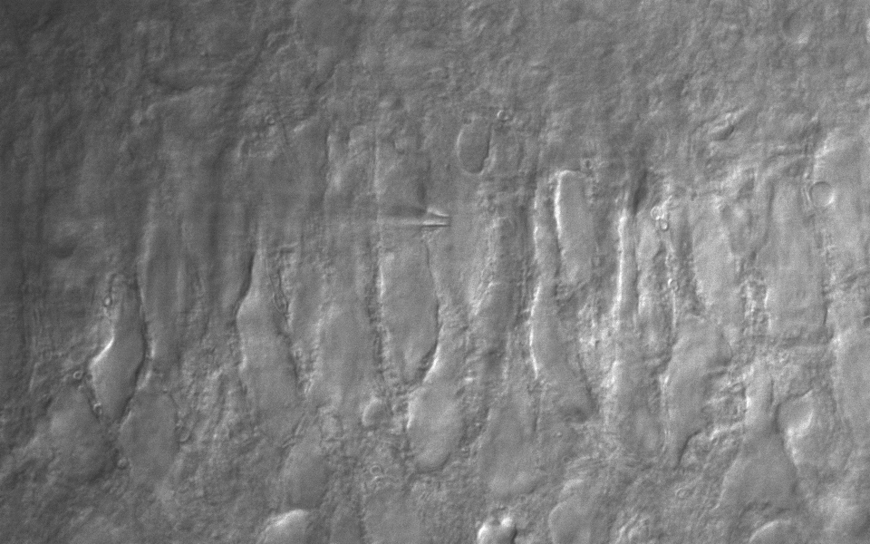
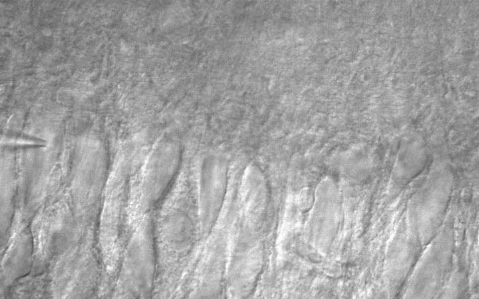
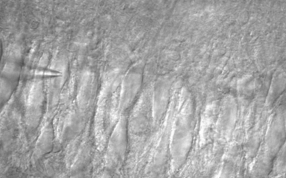
Available measuring height with additional unit, JNO-MHU
※ JNO-MHU is sold separately as optional unit.
Left: Focus on top of sample (Z-axis reset), Right: Focus on bottom of sample (Z-axis height measurement)
< Measuring condition : over 20x objective, temp 20°C >
JNO-MHU is equipment to measure the height of sample, equipped with Z-axis stage handle. It could be equipped easily with new purchasing or existing microscope


