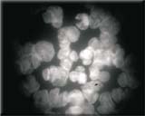
F-view II
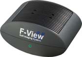
F-View II 는 12-bit monochrome CCD 카메라 입니다.
이 제품은 극도로 높은 감도, 저 노이즈, 고해상도의 정량화할 수 있는 이미지를 요구하는 응용분야에 이상적입니다.
F-View II 는 FireWire 기술 (IEEE1394)에 의한 Peltier 냉각 및 전력을 공급받으며 FISH 혹은 Single band-pass 필터를 사용하는 다중 노출 형광 응용분야를 위해 특별히 설계되었습니다.
F-View II 는 “analySIS Five” Image Analyzer Software에 완전히 통합됩니다.
Digital solutions – Developed to be exceptional!
Soft Imaging System’s digital cameras are designed to meet the steadily rising demands on digital image acquisition for all areas of microscopy. And so is the newly developed 12-bit, cooled CCD color camera F-View II, our latest 12-bit, Peltier-cooled, FireWire™-driven solution for fluorescence microscopy.
The search mode enables you to rapidly locate and conveniently focus in on the area of your sample of interest. This prevents any unnecessary bleaching of the sample.
The camera will automatically switch over to the high-resolution mode for acquisition. F-View II can also be easily operated using a laptop via the FireWire™ port.
All camera functions are fully controlled by the analySIS® image-processing software. Real-time functions enable the use of the entire dynamic range under all conditions and guarantee the best contrast.
F-View II’s integration into analySIS® provides all the capabilities and advantages of state-of-the-art digital image processing and analysis, ranging from image labeling, archiving, report generation and e-mailing, as well as photo-realistic printouts without the darkroom.
The CCD chip of the F-View II has 1376 x 1032 pixel resolution. The 12-bit dynamic range means that even acquisition of images with very bright and faint regions is dynamically optimal.
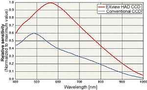
CCD elements are so sensitive that they have no trouble detecting even the weakest of signals. The electronic shutter offers variable exposure times ranging from 100 µs to 160 seconds.
The high speed ADC (Analog-Digital Converter) working at a clock rate of 20 MHz in full 12-bit dynamic range is able to perform double sampling even at a readout rate of 20 MHz. Various frame rates are supported by this camera. For example, the camera can be set to acquire at a high frame rate of more than 22 fps at TV resolution using 2x binning. View your zoomed-in sample, locate the area of interest and focus – all conveniently onscreen. No longer are you forced to trade off speed for quality. For acquisition the system switches automatically into the high-resolution mode. This avoids bleaching of your fluorescence specimen and offers optimal performance when setting parameters.
FireWire™ technology guarantees that the F-View II installation is easy on any PC or laptop equipped with a FireWire™ port. The days when you were limited to a frame grabber and just a single camera are history. FireWire™ technology enables you to use multiple cameras on the same PC.
Image noise is generally the result of the CCD chip not being cooled to a low enough temperature (“dark current”). For the F-View II, thermal noise is not a problem because the CCD chip is Peltier-cooled and stabilized at 10°C. Noise is further suppressed by the application of a highly efficient digital readout technique known as Correlated Double Sampling (CDS).
With its elegantly compact, newly designed housing, the F-View II can be mounted onto all light microscopes with a C-mount adaptor. No other interfaces or adaptors are necessary. Plus, you only need one cable for getting data and power to the PC’s FireWire™ port. No more clutter and no “octopus” of cables getting in your way.
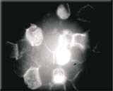
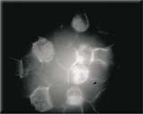
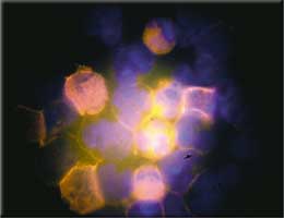
DAPI, PE, APC, PerCP & FITC image acquisitions
of stem cells: combined acquisitions done via themFip analySIS® add-in
source: Dr. Hans Wessels
University Clinic for Children, Tübingen, Germany
F-View II is fast. Put this in combination with the high speed of today’s CPU’s, and you’ve got an attractively broad range of real-time functions within analySIS®available to you. These include automatic contrast control, sharpness monitoring and histogram display.
A vast library of text, graphic and editing functions is available for labeling images. Special filters and professional particle analysis assist in more extensive investigation of images. All this makes it simple to obtain reliable and reproducible results quickly.
Photo-quality printouts can be obtained following acquisition without any need for a darkroom nor its developing chemicals. And these photo-quality printouts are in your hands in minutes.
|
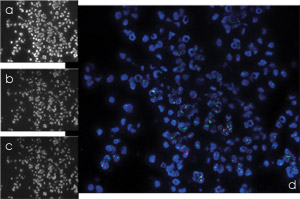
a) Her2 gene (Spectrum Orange)
b) DNA (DAPI) c) Chromosome (Spectrum Green) d) Combination with mFip |
Specimen: Norbert Wey
University of Zürich Department of Pathology Switzerland |
Archiving
analySIS® offers you a powerful, fully networkable, image-archiving system that handles all the images and data generated during the process of image acquisition and documentation. Images can be stored along with text, sheets and other graphics as records for complete documentation of tasks and processing steps. Input, display and query masks can be defined independently.
Automatic report generation
Now you can produce multi-page reports quickly and efficiently. Select multiple images in the image database and insert them all into the report via a single command. In addition to the images themselves, you can have information from any database field automatically included in reports. Automatic scaling, detail zooms, and more – all available to optimize the way you work with images.
All documents generated using the analySIS® software can be inserted into reports. Use the report generator to print out images, related measurement sheets, and diagrams – all on the same page. This report generator provides you with the utmost flexibility for page layout and design. You set up your own templates exactly the way you want them to be. Templates need to be created just once. Templates are what your reports are based on and ensure that the appearance of your documents is uniform. Use the RTF Export function to export your reports to MS Word for continued editing.
Software control of all camera functions
Depending on options, analySIS® expansion level and
![]() Real-time automatic contrast control
Real-time automatic contrast control
![]() Real-time automatic white balance
Real-time automatic white balance
![]() Real-time shading correction
Real-time shading correction
![]() Real-time sharpness monitor
Real-time sharpness monitor
![]() Real-time histogram display
Real-time histogram display
![]() Real-time FFT computation and display
Real-time FFT computation and display
Specifications
| Image Device | 2/3 inch monochrome CCD Sensor |
| (Effective area 8.9 x 6.7 mm array) | |
| Effective Pixels | 1376 x 1032 pixel, 6.45 um square pixels |
| Frame Rate | > 22 fps @ 2x binning |
| 39 fps @ 4x binning | |
| 72 fps @ 8x binning | |
| Binning | 2x, 4x, 8x |
| Dynamic Range | 12 bit |
| Exposure | 100 us ~ 160 sec |
| Cooling | Peltier cooled, 10℃ @ 25℃ ambient |
| Pixel clock rate | 20 MHz |
| Readout noise | < 2 counts |
| Non-linearity | < 0.6% |
| Anti-blooming | > 300 |
| Dimensions (W x H x D) | 100 x 85 x 50 mm |
| Mass | 570g |
| Temperature monitor | CCD chip & housing |
| Temperature stabilized | Yes, ± 0.5 ℃ |
| Interface Connector | FireWire (IEEE1394) |
| Lens mount | Standard C-mount |

